EJCRIM 2023 CiteScore
| 2.1 = | 1.730 Cit. to date |
| 842 Docs. to date |
Last updated on 05 March, 2024
Updated monthly
Updated monthly
Powered by


|
Views: 4494
HTML: 325
PDF: 577
|
Objective: To describe hypocalcaemia due to vitamin D deficiency in ‘hikikomori’ syndrome.
Materials and methods: A 37-year-old man with ‘hikikomori’ syndrome for a year was admitted with hypocalcaemia (serum ionic calcium 1.17 mmol/l). Serum 1,25(OH)2-vitamin D3 determined by liquid chromatography–tandem mass spectrometry was depressed at 12.1 pg/ml (29.0 pmol/l) and plasma intact PTH elevated at 324 ng/l. Administration of 1 ?g/day 1?(OH)-vitamin D3 and 1 g/day calcium lactate for 1 week normalized calcium and PTH, and raised 1,25(OH)2-vitamin D3 to low normal levels.
Conclusion: This is the first report of hypocalcaemia due to vitamin D deficiency in a patient with ‘hikikomori’ syndrome.
|
Views: 1047
HTML: 647
PDF: 453
Figure 1: 0
|
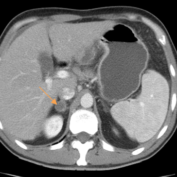
Addison’s disease is an endocrine disorder characterized by primary adrenal insufficiency due to various causes. Mycobacterium tuberculosis infection was a major cause in the past but is rare nowadays. We describe a patient admitted to our hospital who was diagnosed with tuberculous Addison’s disease.
|
Views: 1101
HTML: 1002
PDF: 434
Figure 1: 0
Figure 4: 0
Figure 2: 0
Figure 3: 0
|
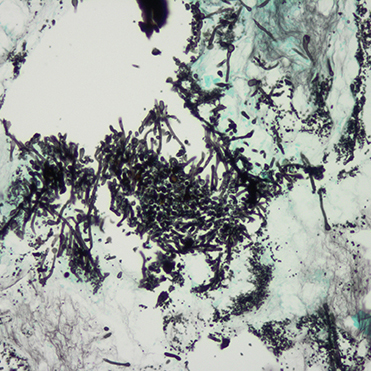
Candida infection of the gastrointestinal (GI) tract is rare in the immunocompetent individual. In immunocompromised patients, it frequently involves the oesophagus, but extra-oesophageal involvement is uncommon. We report a case of primary and isolated gastroduodenal candidiasis. Upper GI endoscopy with biopsy of gastric mucosa was crucial for making the diagnosis. The patient showed transient improvement after therapy with fluconazole.
|
Views: 1168
HTML: 255
PDF: 438
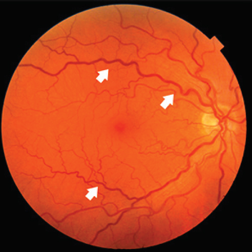
Retinal vessel tortuosity: 0
Dolichoectasia of the vertebrobasilar artery: 0
|
Anderson-Fabry’s disease (AFD) is a rare, X-linked lysosomal storage disorder caused by the complete deficiency or attenuated activity of the enzyme ?-galactosidase A, leading to progressive systemic intracellular accumulation of glycosphingolipids and subsequent cellular dysfunction, inflammation and fibrosis. Fever is a frequently misinterpreted symptom in the early stages of the disease, leading to diagnostic delay. We present the case of a 35-year-old man admitted to our Periodic Fever Research Centre for long-lasting recurrent episodes of fever of unknown origin. After extensive assessment, we diagnosed AFD associated with a novel GLA mutation. We started enzyme replacement therapy with clinical benefit and complete remission of fever.
|
Views: 1784
HTML: 295
PDF: 500
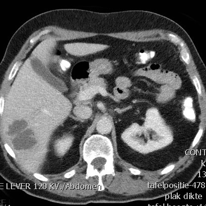
Figure 1: Polyp in the hepatic flexure: 0
Figure 2: CT-scan showing the liver abscess: 0
|
We present the case of a patient who developed a liver abscess following screening colonoscopy. A colorectal screening program was introduced in the Netherlands in 2014 in order to reduce mortality from colorectal cancer. The patient in this report, a 63-year-old man with no significant medical history, underwent polypectomy of two polyps. Four days afterwards he presented to our emergency department with fever, nausea and vomiting. He was diagnosed with a Klebsiella pneumoniae liver abscess and was successfully treated with antibiotics for 6 weeks. This case highlights one of the risks of screening colonoscopy. Given the high number of colonoscopies due to the colorectal screening programs, we should be aware of complications in this mostly asymptomatic group of patients.
|
Views: 1329
HTML: 4879
PDF: 452
Figure 1: 0
Figure 2: 0
Full Reference List: 0
|
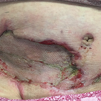
A patient with post-Cesarean wound complication was treated for necrotizing fasciitis (NF) with sharp debridement and broad-spectrum antibiotics. Several operations and three weeks later, her abdominal skin, subcutaneous fat, right-sided rectus abdominus, and underlying fascia had been removed without any improvement in granulation tissue. Original pathology samples demonstrated sheets of necrosis consistent with NF, but were re-reviewed by a dermatopathologist who diagnosed the patient with pyoderma gangrenosum (PG). She was started on high-dose steroids and dapsone, and her wound quickly showed signs of improvement. Anchor bias delayed the initiation of steroids and diagnosis of PG as the surgical, medical, and consulting teams were hesitant to stray from the diagnosis of NF.
| 2.1 = | 1.730 Cit. to date |
| 842 Docs. to date |


Publisher
Official Journal of the
European Federation of Internal Medicine
www.efim.org
Publisher: SMC media Srl
Via Giovenale, 7 - 20136 Milan - Italy
P.IVA 07626490960
info@ejcrim.com
www.ejcrim.com - ISSN: 2284-2594 - © EFIM 2014-2023, Published by SMC Media srl, Italy - Privacy policy

