ABSTRACT
Listeria monocytogenes is a gram positive bacillus responsible for listeriosis. This infection manifests mainly as bacteremia and / or central nervous system (CNS) infection. Involvement of other sites is rare. Pleural listeriosis is an uncommon presentation of L. monocytogenes infection and there is little data to guide the management of these patients. First-line antibiotics used empirically to treat bacterial respiratory infections are ineffective in treating these L. monocytogenes infections, which contributes to the progression of the infection and a worse prognosis.
We present a case report of a patient admitted to an intensive care unit with septic shock secondary to systemic listeriosis with L. monocytogenes isolation in pleural fluid culture and blood cultures. The evolution of the hospitalization and the clinical outcome are reported.
LEARNING POINTS
- Pleural listeriosis is an infection rarely observed in clinical practice.
- It is typically found in immunosuppressed patients.
- Delay in the identification of this microorganism and the empirical use of inappropriate antibiotics may lead to an adverse outcome.
KEYWORDS
Listeria monocytogenes, pleural listeriosis, immunosuppression
INTRODUCTION
Cases of listeriosis are uncommon, mainly affecting patients in the neonatal period, pregnant women, people over 60 and immunosuppressed at any age. The most common forms of presentation in adults are meningitis and / or encephalitis and sepsis, with few cases of listeriosis in other locations. Pleural listeriosis is a rare manifestation of L. monocytogenes that is found primarily in immunosuppressed adult. In literature, most cases are described in patients with malignant disease. We describe the case of a 69-year-old patient admitted to an intensive care unit (ICU) for septic shock with identification of L. monocytogenes in pleural fluid and blood cultures.
CASE PRESENTATION
TWe present a case report of a 69-year-old patient with primary arterial hypertension, dyslipidemia, ischemic heart disease with severe heart ejection fraction 15-20% and a mechanical aortic valve, stage 4 chronic kidney disease and early-stage Alzheimer's dementia. This patient was a middle-class art teacher living in an urban environment, with no history of recent travel or dairy intake.
The patient was admitted to the emergency department with non-bloody diarrhea for the last four days, diffuse abdominal pain, dyspnoea and unquantified fever. On admission, the patient was prostrated and hypotensive. Chest X-ray revealed a homogeneous opacity involving the middle and lower lobes of the right hemithorax, suggestive of pleural effusion (Fig. 1). Analytically, to be highlighted, protein C-reactive protein was 27 mg/dL, serum creatinine was 6 mg/dl and the arterial gas analysis demonstrated a hypoxemic respiratory failure. Lactate level at admission was 1,37 mmol/L. Septic shock was assumed, with multiorgan dysfunction, and empirical antibiotic therapy with ceftriaxone was started. He was admitted to the ICU. Despite fluid filling, the patient required vasopressor support, invasive mechanical ventilation, and continuous renal function replacement technique and was transferred to the ICU. Due to the severity of the condition, it was decided to early escalate antibiotic therapy for meropenem and linezolid. Later, L. monocytogenesis was identified both in blood cultures and in the pleural fluid. HIV was negative, and due to the clinical condition of the patient, it was decided to delay the invasive diagnostic investigation of possible causes of immunosuppression. After identifying the causal microorganism, more epidemiologic questions were asked. The family denied a history of consumption of contaminated food and no additional cases suggestive of listeria infection were identified.
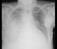
Figure 1 (click to enlarge)
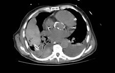
Figure 2 (click to enlarge)
Figure 1. Chest x-ray showing homogeneous opacity involving the middle and lower lobes of the right hemithorax, suggestive of pleural effusion
Figure 2. Chest CT scan documents an extensive multiloculated right pleural effusion
The chest CT scan revealed extensive multiloculated right pleural effusion (Fig. 2). In this context an attempt was made for pleural decortication which was complicated with hemothorax. Linezolid was changed to ampicillin, maintaining the already established meropenem, with a decrease in inflammatory parameters. The patient was treated with a total of 12 days of meropenem and 21 days of ampicillin, always in meningeal doses despite the absence of evidence of CNS involvement. However, on the 13th day of ampicillin, due to a further increase in inflammatory parameters and worsening of pleural imaging, piperacillin-tazobactam was added. The patient evolved with new laboratory and clinical worsening. Given the co-morbidities and the worsening clinical picture, it was decided to favor patient comfort and suspension of invasive measures at day 25 of admission. The patient died after a month of ICU stay.
DISCUSSION
As in the rest of the world, listeriosis is an uncommon zoonosis in Portugal. It is caused by an optional sporulated, anaerobic gram-positive intracellular bacillus called L. monocytogenes [4, 5]. In adults, transmission of this bacillus occurs through contaminated food [2,4,5]. From there, it can spread to any part of the organism, yet it presents a particular tropism to the CNS [3]. This bacterium is recognized as an opportunistic microorganism< sup>[1] since L. monocytogenes infection is related to an individual's cellular immunodepression status and is a rare cause of pathology in healthy patients [3,5]. It affects predominantly pregnant women, newborns, individuals over 60 and immunocompromised young adults. Clinically, in individuals over the age of 60 and in immunocompromised patients of any age, sepsis and CNS infection predominate, namely meningitis and encephalitis [1]. These situations are the most serious manifestations of listeriosis.
First-line treatment for listeriosis is ampicillin, whether or not associated with gentamicin in severe cases. An alternative is trimethoprim sulfametazole [5]. In all cases of invasive listeriosis, meningitis doses of antibiotics should be used, even without evidence of CNS attainment, due to the high affinity of these microorganisms to this location. The duration of treatment varies from 2 to 6 weeks according to the severity of the clinical condition and CNS involvement[5]. Infection of extra-CNS locations with L. monocytogenesis, although rare, should be part of our differential diagnoses when facing an immunosuppressed individual [1,3,4,5]. However, this infection is usually only considered when there is clear CNS attainment. There are few cases of pleural listeriosis described in the literature, so there are few guidelines regarding the treatment of patients with respiratory infections caused by this microorganism[2]. Given the limited experience, the optimal duration of treatment for pleural infection by L. monocytogenes is unknown, but a minimum duration of 2 weeks is accepted and the first-line choice is the combination of ampicillin and an aminoglucoside, such as gentamycin [4, 5].
Since L. monocytogenes is rarely the causative agent of respiratory infections, this bacterium is generally not considered as an etiological agent in the initial differential diagnosis of this type of infection [1, 3, 4]. On the other hand, it is a slow growing microorganism and cultures may take up to 5 to 7 days to identify this microorganism. This may lead to the empirical use of ineffective antibiotics in the treatment of respiratory infections by L. monocytogenes such as cephalosporins [4]. This promotes determines a worse prognosis. Respiratory infections caused by L. monocytogenes reveal a great vulnerability of the individual and are associated with more invasive forms and high morbidity and mortality [2]. With the exception of benign manifestations of self-limiting gastroenteritis in healthy patients, the identification of this microorganism should motivate the study of causes of immunosuppression [5]. In this case, no investigation was performed due to the lack of benefit determined by a poor clinical course.