ABSTRACT
Very often in clinical practice, an inflamed pelvic appendix shows left lower quadrant abdominal pain as the primary painful area. The clinicians are anchored to the most prominent symptom, thereby taking an unnecessary detour in reaching an accurate diagnosis.
A 40-year-old man presented to our emergency department with persistent lower left abdominal pain with a fever of 38 oC from a day earlier. He had a good appetite and repeatedly complained of severe constipation at the time of his visit. Physical examination revealed tenderness in the lower left abdomen without a peritoneal sign. Abdominal ultrasound and non-contrast-enhanced computed tomography revealed a left hydroureter. The next day, a radiologist pointed out the possibility of appendicitis. An urgent laparoscopic appendectomy was performed.
The intriguing point of this case is the diagnostic delay because of three anchoring biases. First, the typical right lower abdominal pain of appendicitis was shielded by the intense left lower abdominal pain. Moreover, the presence of a left hydroureter distracted the physicians from the actual location of the pain. Furthermore, the presence of constipation anchored the physicians to constipation as the cause of abdominal pain.
In overcoming these biases, specific diagnostic strategies to avoid biases should be implemented.
LEARNING POINTS
- If a patient has unexplained lower left abdominal pain, it is advisable to deploy a “searchlight” strategy.
- When a hydroureter was found to have no apparent source obstruction, a vertical tracing strategy should have been undertaken to detect its root cause.
- To avoid the wrong diagnosis through anchoring bias, pivot and cluster strategy – deploying differential diagnosis specific to the initial diagnosis (constipation in this case) – should be adopted at the start, considering the important differential diagnosis and thus preventing a missed diagnosis.
KEYWORDS
Appendicitis, debiasing, anchoring, lower abdominal pain, appendectomy
INTRODUCTION
Very often in clinical practice, an inflamed pelvic appendix shows left lower quadrant abdominal pain as the primary painful area. The clinicians are anchored to the most prominent symptom, thereby taking an unnecessary detour in reaching an accurate diagnosis. This report describes a patient diagnosed with appendicitis with persistent lower left abdominal pain. The intriguing point of this case is the diagnostic delay because of three anchoring biases. The diagnosis of appendicitis is sometimes challenging. In overcoming these biases, specific diagnostic strategies to avoid biases should be implemented.
CASE DESCRIPTION
A 40-year-old man presented to our emergency department with two weeks of persistent lower abdominal pain. It was a continuous pain of constant intensity. He presented with a history of fever of 38°C since the previous day, constipation after the onset of pain, good appetite and no vomiting or diarrhoea. He repeatedly complained of severe constipation when he reported to us. He had a medical history of depression.
On examination, his temperature was 38.2°C, respiratory rate was 15 breaths per minute, blood pressure was 128/86 mm Hg and pulse was 75 beats per minute. Physical examination revealed tenderness in the lower left abdomen without a peritoneal sign. The other physical findings were normal.
Laboratory evaluation revealed an elevated white blood cell count of 12.17×10⁹/L (neutrophils: 82.6%) and C-reactive protein concentration (11.97 mg/dL). Liver and kidney functions were within normal range. Abdominal ultrasound and non-contrast-enhanced computed tomography (CT) revealed a left hydroureter (Fig. 1). Based on the imaging findings, abdominal pain caused by hydronephrosis was suspected, and he was discharged home.
The next day, a radiologist pointed out the possibility of appendicitis (Fig. 2), and the patient was recalled to the hospital. On further inquiry, the patient admitted a vague lower abdominal pain, which was not so prominent around the appendix area. Further abdominal examination confirmed mild pain at the appendix base and left lower abdominal tenderness.
An urgent laparoscopic appendectomy was performed, revealing appendicitis with partial abscess formation. The postoperative course was uneventful, and the patient was successfully discharged home.
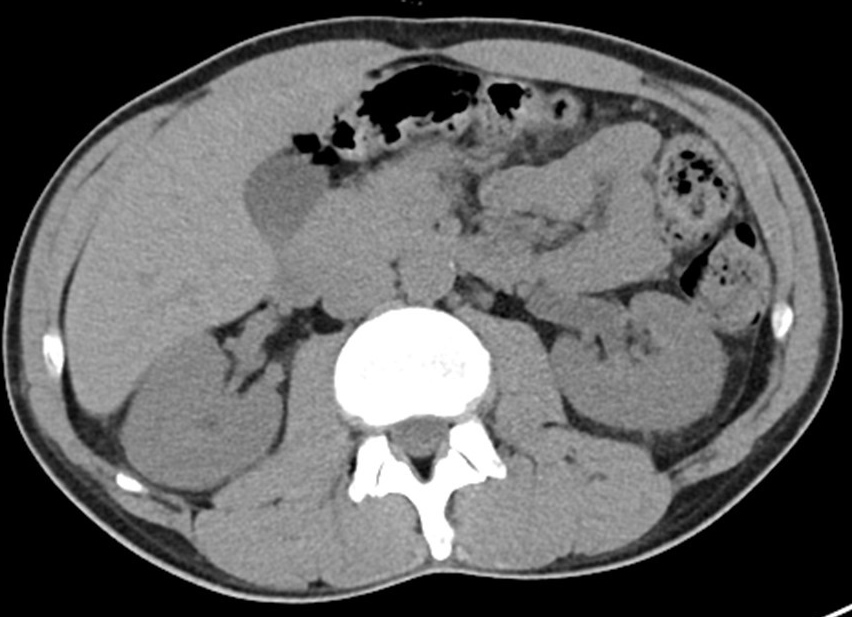
Figure 1 (click to enlarge)
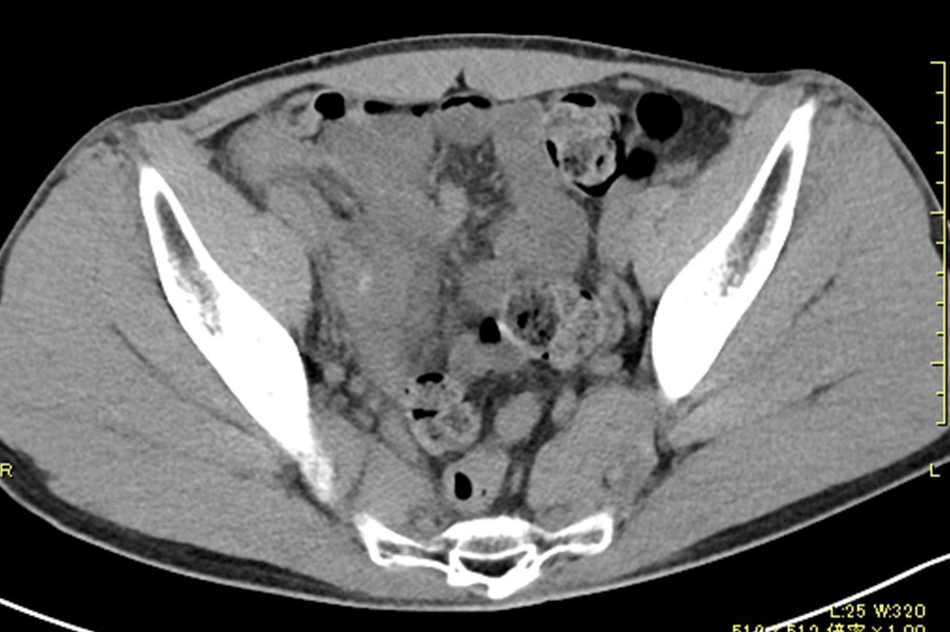
Figure 2 (click to enlarge)
Figure 1. Non-contrast-enhanced computed tomography (CT) revealed a left hydroureter.
Figure 2. Non-contrast-enhanced computed tomography (CT) revealed the possibility of appendicitis.
DISCUSSION
Three anchoring biases contributed to the diagnostic delay in this case. First, the typical right lower abdominal pain of appendicitis was shielded by an intense left lower abdominal pain, making it difficult to diagnose. The clinicians were anchored to the most prominent symptom of the left lower abdominal pain, thereby taking an unnecessary detour in reaching the accurate diagnosis. Second, anchoring to the presence of left hydroureter as the most prominent finding on CT distracted the physicians from the location of the pain. Third, the presence of constipation as a conspicuous symptom in this patient led the physicians to believe that constipation was the sole cause of the abdominal pain.
If the appendix is positioned posteriorly without contact with the anterior peritoneum, the patient may not show significant local tenderness in the right lower abdomen[1]. It was reported that the appendix is located in the pelvis in about 15% of cases [2]. Furthermore, in real-world clinical practice, an inflamed pelvic appendix is often characterised by pain in the left lower quadrant abdominal area. In our patient, there was prominent left lower abdominal pain without any right lower abdominal pain. An article regarding the location of the appendix base in relation to the McBurney’s point reported that the appendix base was more than 10 cm away from the McBurney’s point in 15% of cases, and if such cases affected the south-east quadrant, left lower spontaneous pain and tenderness may occur [3].
Appendicitis would otherwise also have presented with the same symptoms as in this case with the reported position of the appendix; however, the patient did have mild pain at the base of the appendix. Pain around the appendix base may be present even in atypical appendicitis cases. Therefore, a careful examination of the appendix base is significant to indicate appendicitis. Consequently, if a patient has unexplained lower left abdominal pain, it is advisable to deploy a “searchlight” strategy. In this strategy, doctors concentrate on palpating the abdomen, shifting the palpating location inch by inch as if they were focusing a searchlight on each area in sequence. This extensive search makes it possible to discover a finding that may otherwise have been overlooked. This strategy may make it possible to detect latent lesions in addition to those already known, thereby identifying the tenderness at the appendix base, similar to the present case.
Another anchoring bias observed in this case was the presence of a dilated left ureter on CT, which was attributed to being associated with the left lower abdominal pain. This anchoring bias boils down to availability bias, a physicians’ heuristic whereby they associate left ureteral dilation with left lower abdominal pain. One possible reason for the presence of a hydroureter in this patient was that the inflammation of the appendix in the pelvis extended to the periureter area, resulting in ureteral stenosis. When the physicians had suspected this possibility on studying the CT, they should have continued to explore the main locus of inflammation and avoided being trapped by the availability bias. When they saw a hydroureter in which no apparent source obstruction was found, a vertical tracing strategy, an endeavour to detect its root cause, should have been undertaken [4].
Physicians treating abdominal pain and constipation patients have been reported to be more likely to miss appendicitis in the emergency department [5]. In the present case, the patient’s history may have led to anchoring that the abdominal pain was probably due to constipation, causing “hustle bias” and early closure. To avoid the wrong diagnosis through anchoring bias, pivot and cluster strategy[6] – deploying differential diagnosis specific to the initial diagnosis (constipation in this case) – should be adopted at the start, considering the important differential diagnosis and thus preventing a missed diagnosis.
CONCLUSION
It is essential to make an effort to clarify the primary locus of inflammation with careful physical findings and imaging readings based on strategies that are not confounded by inherent biases. Verifying the validity of the diagnosis over time will further ensure an accurate diagnosis.
The diagnosis of appendicitis is sometimes challenging. In this case, the patient presented with atypical appendicitis, and triple anchoring made it challenging for the physicians to reach an accurate diagnosis. To overcome these biases, specific diagnostic strategies to avoid biases should be implemented.