ABSTRACT
Purpura fulminans (PF) is a dermatological manifestation of a life-threatening condition characterized by disseminated intravascular coagulation and endovascular thrombosis. The idiopathic/infectious form is the most common and usually associated with infection by Neisseria meningitidis or Streptococcus pneumoniae. We describe a case of Morganella morganii-induced bacteriaemia complicated with PF in an individual who had undergone a recent urinary tract infection intervention. The patient presented with purpuric skin lesions, fever and hypotension but had no alterations in coagulation parameters or disseminated intravascular coagulation. Aggressive early resuscitation, intravenous antibiotics and wound care were essential to a favourable response.
LEARNING POINTS
- Purpura fulminans is a dermatological manifestation of an underlying life-threatening condition, and is characterized by disseminated intravascular coagulation and skin necrosis.
- It is a morbid and potentially fatal condition that can be a cutaneous manifestation of Morganella morganii bacteraemia.
- Early identification and accurate diagnosis of the underlying cause can help minimize morbidity and mortality; management should be tailored to the individual, with the use of intravenous antibiotics, necrotic skin excision and aggressive early resuscitation.
KEYWORDS
Purpura fulminans, Morganella morganii
INTRODUCTION
Purpura fulminans (PF) is a medical emergency with high mortality, requiring rapid diagnosis and treatment. It is a rare syndrome characterized by disseminated intravascular coagulation and endovascular thrombosis which results in cutaneous purpura. There are three types: neonatal, idiopathic and infectious. The latter is the most common form, caused in most cases by Neisseria meningitidis infection in children. However, other agents can also cause this condition, mainly Gram-negative bacteria. Nevertheless, an association between this entity and urinary tract infection caused by Morganella morganii has not previously been reported.
CASE DESCRIPTION
We present the case of an 80-year-old man brought to the emergency department (ED) after being found unconscious on his doorstep. He had a history of laryngeal neoplasia, arterial hypertension, dyslipidaemia and vulgar psoriasis which mainly affected his lower extremities.
He had been taking aspirin, allopurinol, enalapril and lercanidipine once a day for several years. He had not started any new medication, had no allergies and was not taking anticoagulants or antibiotics. However, he had undergone a cystoscopy about 7 days before presenting to the ED.
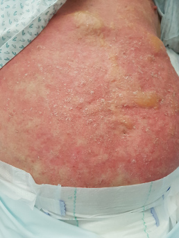
Figure 1 (click to enlarge)
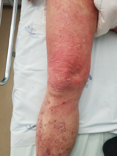
Figure 2 (click to enlarge)
Figure 1. Blistering lesions with citrine content and detachment all over the abdomen
Figure 2. Blistering lesions with citrine content and detachment all over the lower limb mixed with vulgar psoriasis lesions
The laboratory study showed AKIN 1 acute kidney injury (creatinine of 1.98 mg/dl) and elevated C-reactive protein (21 mg/dl). The blood count and clotting tests were within normal limits, including D-dimers (270 ng/ml; reference values 0–300 ng/ml) and fibrinogen 430 mg/dl (reference values 200–500 mg/dl). Urinary sediment was active with leukocyturia (>200/field) and proteinuria (250 mg/dl).
We admitted the possible diagnosis of sepsis with a urinary source and started ceftriaxone and vancomycin after collecting cultures. Supportive therapy with antipyretics and fluids was administered with a good response.
A skin biopsy showed necrosis of the entire thickness of the epidermis, underlying which the capillaries were markedly congestive and thrombosed, with extravasation of red blood cells and inflammatory infiltrate. The findings were compatible with a vaso-occlusive syndrome of small vessels on the PF spectrum.
The urine culture result was negative, but wound exudate culture and blood cultures isolated M. morganii with resistance to penicillin. We changed to gentamicin, which the patient took for 10 days, with a good response; wound care was performed with permanganate patches. He was discharged after 15 days, and at the follow-up visit 3 months later, the skin lesions had practically healed.
DISCUSSION
PF is a haemorrhagic necrosis and microvascular thrombosis syndrome. It is characterized by disseminated microvascular thrombi that cause extravasation of red blood cells, haemorrhagic skin infarction, and peripheral gangrene. There are three types, but the most common form, idiopathic or acquired, usually occurs about 7–10 days after sepsis due to Gram-negative endotoxin-producing bacteria, such as N. meningitidis and Streptococcus pneumoniae. However, other endotoxin-producing Gram-negative bacteria can also cause this condition [1].
M. morganii is a Gram-negative bacillus that colonizes the mucosa of the gastrointestinal tract of humans and animals. Typically, it is a nosocomial pathogen and does not produce endotoxins [2]. It is responsible for infections in the urinary tract and postoperative wounds, mainly in immunosuppressed patients. There are few studies on Morganella bacteraemia, but patients with sepsis have more comorbidities and worse sepsis. There is an increased risk of inappropriate empirical treatment and extended hospitalization, and a higher death rate. The main risk factors are advanced age, severe underlying disease, hospitalization, recent surgery, and concomitant use of antibiotics. The mortality rate with bacteraemia was 22–38% [3]. In the case presented, the patient had undergone a urinary intervention 7 days before developing the condition, which provided an opportunity for bacteraemia to develop.
In a patient with bacterial sepsis, both vasculitis and thrombotic components may be present [4]. There are several publications about PF, and despite the devastating clinical course associated with this haemostatic complication of infection, the mechanism of the condition remains poorly understood. Severe acquired protein C deficiency and dysfunction of the protein C-thrombomodulin pathway and of other systems that exert a negative regulatory effect on coagulation have been implicated. However, a definite physiopathological mechanism has not been identified [5].
This report is the first publication to describe bacteraemia by M. morganii triggering a vasculitic response in the form of PF. As the exact mechanism underlying this condition is unknown, further studies are required to identify the cause.
One of the diagnoses to exclude is disseminated intravascular coagulation (DIC) [1], which involves the abnormal and excessive formation of thrombin and fibrin, with increased platelet aggregation and consumption of coagulation factors. However, we did not detect thrombocytopenia, prolongation of partial thromboplastin time and prothrombin time, increased concentration of D-dimers or decreased fibrinogen. Proteins C and S were within normal limits.
Although the mortality associated with this type of infection is high, targeted antibiotic therapy and appropriate supportive therapy allowed for adequate treatment of the patient, preventing multiorgan failure. To reduce mortality, early recognition and treatment of PF with supportive therapy, implementation of aggressive resuscitation and administration of antibiotics, is essential.
CONCLUSION
PF is a potentially fatal condition that can be a cutaneous manifestation of M. morganii bacteriaemia. Early recognition and accurate identification of the underlying cause can reduce fatal complications. Moreover, in our case, aggressive early resuscitation, intravenous antibiotics, correction of coagulopathy, and wound care were essential to a favourable response.