ABSTRACT
Coxiella burnetii causes Q fever, which is found worldwide and can be acute or chronic. This case report describes a 72-year-old man whose bilateral lower limb pain revealed a paravertebral abscess at L2–L3 due to Q fever spondylodiscitis. Surgical drainage of the abscess was performed and medical treatment is ongoing.
Q fever is endemic in Portugal and transmitted by inhalation of aerosols containing spores from infected animals (cattle, goats and sheep) or by ingesting cottage cheese or unpasteurized milk. It has an incubation period of 2–3 weeks and 60% of patients are asymptomatic with only 2% needing hospitalization. Primary infection can manifest in any organ and most cases are self-limiting (self-limited febrile illness, atypical pneumonia or acute hepatitis). Less than 1% of cases evolve to chronic disease, presenting as osteomyelitis or endocarditis.
Chronic disease poses a diagnostic challenge and spondylodiscitis has an insidious evolution. Diagnosis requires microbiological and clinical confirmation. Serological and polymerase chain reaction tests are used for diagnosis.
Acute disease is usually treated with doxycycline for 3 weeks to avoid evolution to chronic disease. Chronic disease requires 18–24 months of doxycycline with hydroxychloroquine. Acute disease can recur so follow-up is essential as chronic Q fever can result in morbidity and mortality. In Portugal Q fever is a notifiable disease due to the epidemiological risk.
LEARNING POINTS
- Coxiella burnetii spondylodiscitis is rare so clinicians should be aware of it.
- The diagnosis of Q fever is challenging, especially chronic disease that presents with endocarditis, osteomyelitis or spondylodiscitis.
- Spondylodiscitis treatment is particularly challenging and may need neurosurgical intervention.
KEYWORDS
Coxiella burnetii, Q fever, spondylodiscitis
INTRODUCTION
Coxiella burnetii is a Gram-negative intracellular bacterium which causes Q fever, a globally distributed zoonosis which can present with acute and chronic manifestations [1].
Q fever is endemic in Portugal and a notifiable disease given the epidemiological risk it poses [1]. It is transmitted by inhalation of aerosols containing spores from infected animals (main reservoir: cattle, goats and sheep) or by ingesting cottage cheese or unpasteurized milk [1].
It has an incubation period of 2–3 weeks and 60% of patients are asymptomatic [2]. Primary infection can manifest in any organ and most cases are self-limiting (self-limited febrile illness, atypical pneumonia or acute hepatitis). Less than 1% of cases evolve to chronic disease and present as bone infection [2].
Chronic osteomyelitis in Q fever has an insidious evolution, presents histological evidence of granulomatous inflammation, and lacks evidence of the presence of alternate microorganisms, including mycobacteria [3].
Diagnostic reference methods include serological and polymerase chain reaction tests [4]. The disease can recur so follow-up is essential as chronic Q fever and its inflammatory syndrome can lead to morbidity and mortality [5].
Treatment includes doxycycline and other regimens including quinolones [4]. Given the challenges of vertebral pyogenic infection, surgical drainage presents additional difficulties [3].
CASE DESCRIPTION
A hypertense 72-year-old man was evaluated in the Infectious Disease Unit after orthopaedic evaluation for loss of strength in his lower limbs. The CT scan revealed an inflammatory lesion on lumbar vertebrae L2–L3 and a paravertebral abscess (Fig. 1). The patient lives in a rural setting, has contact with goats, and had a 1-year history of pain measuring 4/10 on the numerical scale with an inflammatory pattern. There were no relevant clinical or physical findings.
The patient was admitted, microbiological samples collected, and a CT-guided biopsy of the lesion performed. Empirical therapy with ceftriaxone and vancomycin was started, but on the 11th day the patient presented generalized macular exanthema on the neck, thorax, abdomen and thighs, which disappeared on digital pressure, fever and cytolysis that resolved with the suspension of antibiotic therapy. Biopsy results were insufficient for diagnosis (only musculoskeletal and fat tissue). Levofloxacin was started and suspended due to vasculitis, so meropenem was instituted empirically while microbiological studies were still negative or pending.
Serological testing revealed C. burnetii antibody titres of 1/512 IgG phase I and 1/128 IgG phase II, and were IgM negative. Blood cultures were negative and the patient remained asymptomatic. Echocardiography ruled out endocarditis. Additional studies were negative (Mycobacterium tuberculosis, Brucella and HIV). Serological testing after 8 weeks showed 1/2048 IgG phase I and phase II titres, confirming the Q fever hypothesis. Treatment was adjusted to doxycycline and hydroxychloroquine. MRI imaging revealed advanced L2–L3 spondylodiscitis and empyema with a left psoas major ipsilateral abscess, so CT-guided drainage was performed. Culture of the drainage fluid was negative.
There was minor clinical improvement given the presence of vertebral destruction and empyema, so neurosurgical evaluation recommended surgical fixation of the vertebrae and drainage of the intracanal empyema, which was successful (Fig. 2). Culture of pyogenic material was negative.
The patient is currently undergoing physical rehabilitation in a convalescence unit and has another 12 months of antimicrobial therapy (for a total of at least 18 months). A multimodal strategy is in place to treat non-oncological chronic pain and medical follow-up is being maintained.
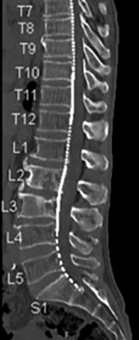
Figure 1 (click to enlarge)
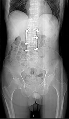
Figure 2 (click to enlarge)
Figure 1. Spondylodiscitis at L2–L3
Figure 2. X-ray after stabilizing surgery
DISCUSSION
Q fever is common in Portugal but frequently undiagnosed [1]. The epidemiological context is important as diagnosis may be difficult if symptoms are mild; disease confirmation is sometimes based only on a chronic presentation and disease severity [1]. However, microbiological criteria (serology) in addition to lesion imaging have been used for diagnosis [4]. Serological tests take time but other microbiological methods may not be as helpful as blood and aspirate (three lesion biopsies) cultures were negative in our patient. The differential diagnosis of inflammatory syndrome and chronic pain frequently includes autoimmune disorders. Given the presence of autoantibodies (antinuclear, anti-mitochondrial, anti-smooth muscle, and especially procoagulant anticardiolipin and lupus anticoagulant), the epidemiological and clinical histories are important for differentiating between clinical entities [5]. The therapy of choice is doxycycline, but chronic cases also need the addition of hydroxychloroquine which allows alkalinization of the acidic vacuoles thus disrupting the bacterial environment in monocytes/macrophages [4]. Q fever should be included in the differential diagnosis of vertebral osteomyelitis in endemic settings, in particular if vascular infection is identified. Imaging-guided aspiration and surgical drainage pose additional risks but in complex cases may be the only way to control the source of pyogenic infection [3].