A Case of Fluorine-18 Fluorodeoxyglucose Positron Emission Tomography/Computed Tomography Imaging of Eosinophilic Granulomatosis With Polyangiitis Combined With Non-tuberculous Mycobacterium
  • Juhyung Kim
    Department of Internal Medicine, School of Medicine, Kyungpook National University, Daegu, Korea
  • Man-Hoon Han
    Department of Pathology, School of Medicine, Kyungpook National University, Daegu, Korea
  • Sang Jin Lee
    Department of Internal Medicine, School of Medicine, Kyungpook National University, Daegu, Korea

Keywords

Positron emission tomography/computed tomography, non-tuberculous mycobacterium, eosinophilic granulomatosis with polyangiitis

Abstract

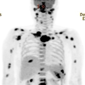
Fluorine-18-fluorodeoxyglucose positron emission tomography/ computed tomography (18FDG-PET/CT) has been used to diagnose vasculitis, tuberculosis and malignancy. Because PET/CT scan show hotspots during an activation of clinically suspected lesions, it is widely used for diagnosis. However, there are rare cases of PET/CT images for vasculitis combined with tuberculosis. Here we report a case of an eosinophilic granulomatosis with polyangiitis in a patient with disseminated non-tuberculosis mycobacterial infection in multiple sites mimicking metastatic malignacy and describe the associated PET/CT scan findings before and after treatment.

VIEW THE ENTIRE ARTICLE

References

  • Lee SJ, Jeong JH, Lee CH, Ahn BC, Eun JS, Kim NR, et al. Development and validation of an (18) F-fluorodeoxyglucose-positron emission tomography with computed tomography-based tool for the evaluation of joint counts and disease activity in patients with rheumatoid arthritis. Arthritis Rheumatol 2019;71:1232–1240.

  • Ito K, Minamimoto R, Yamashita H, Yoshida S, Morooka M, Okasaki M, et al. Evaluation of Wegener’s granulomatosis using 18F-fluorodeoxyglucose positron emission tomography/computed tomography. Ann Nucl Med 2013;27:209–216.

  • Kemna MJ, Vandergheynst F, Vöö S, Blocklet D, Nguyen T, Timmermans SA, et al. Positron emission tomography scanning in anti-neutrophil cytoplasmic antibodies-associated vasculitis. Medicine 2015;94:e747.

  • Soussan M, Abisror N, Abad S, Nunes H, Terrier B, Pop G, et al. FDG-PET/CT in patients with ANCA-associated vasculitis: case-series and literature review. Autoimmun Rev 2014;13:125–131.

  • Nelson DR, Johnson GB, Cartin-Ceba R, Specks U. Characterization of F-18 fluorodeoxyglucose PET/CT in granulomatosis with polyangiitis. Sarcoidosis Vasc Diffuse Lung Dis 2015;32:342–352.

  • Del Giudice G, Bianco A, Cennamo A, Santoro G, Bifulco M, Marzo C, et al. Lung and nodal involvement in nontuberculous mycobacterial disease: PET/CT role. Biomed Res Int 2015;2015:353202.

  • Gioffredi A, Maritati F, Oliva E, Buzio C. Eosinophilic granulomatosis with polyangiitis: an overview. Front Immunol 2014;5:549.

  • Sohn S, Shi HJ, Wang SH, Lee SK, Park SY, Lee JS, et al. Mycobacterium avium complex infection-related immune reconstitution inflammatory syndrome mimicking lymphoma in an human immunodeficiency virus-infected patient. Infect Chemother 2018;50:350–356.

  • van Timmeren MM, Heeringa P, Kallenberg CG. Infectious triggers for vasculitis. Curr Opin Rheumatol 2014;26:416–423.

  • Lin K-H, Wang J-H, Peng N-J. Disseminated nontuberculous mycobacterial infection mimic metastases on PET/CT scan. Clin Nucl Med 2008;33:276–277.
  • Views: 910
    HTML downloads: 90
    PDF downloads: 381


    Published: 2019-11-22
    Issue: Vol 6 No 12 (view)


    How to cite:
    1.
    Kim J, Han M-H, Lee SJ. A Case of Fluorine-18 Fluorodeoxyglucose Positron Emission Tomography/Computed Tomography Imaging of Eosinophilic Granulomatosis With Polyangiitis Combined With Non-tuberculous Mycobacterium. EJCRIM 2019;6 doi:10.12890/2019_001299.