EJCRIM 2023 CiteScore
| 2.1 = | 1.730 Cit. to date |
| 842 Docs. to date |
Last updated on 05 April, 2024
Updated monthly
Updated monthly
Powered by


|
Views: 1469
HTML: 114
PDF: 553
|
Introduction: Phenazopyridine is an over-the-counter urinary analgesic commonly used to alleviate the burning and urgency associated with lower urinary tract infections. Methaemoglobinaemia is an uncommon adverse effect of phenazopyridine use. We report a case of methaemoglobinaemia in a patient prescribed daily phenazopyridine to treat urethral and bladder irritation caused by a chronic indwelling Foley catheter.
Case description: A 55-year-old female resident of a long-term acute care facility with a chronic Foley, tracheostomy and ventilator-dependent respiratory failure was observed to have generalized dusky skin and hypoxia. Pulse oximetry was reading in the high 80s despite administration of 100% FiO2. ABG revealed paO2 of 451, oxyhaemoglobin level 75% and methaemoglobin level 22%. Medication review indicated that the patient was prescribed phenazopyridine 400 mg TID for the previous 2 months. This medication was discontinued. Considering she was chronically taking mirtazapine, she can increase risk of serotonin syndrome should she be administered first-line treatment with methylene blue. Vitamin C was thus instead administered as a second-line agent. Serial ABGs showed a rapid decline in methaemoglobin levels and an increase in oxyhaemoglobin within 2 days.
Discussion: Acquired methaemoglobinaemia is a rare adverse effect of treatment with phenazopyridine. This risk increases when drug dosage and duration exceed manufacturer specifications. Treatment typically includes cessation of the offending drug and administration of methylene blue in severe cases. A thorough medication reconciliation should be performed prior to methylene blue initiation, as patients taking serotonergic medications (for example, MAOIs, SSRIs, SNRIs, TCAs) are at increased risk of serotonin toxicity with co-administration of methylene blue. In these instances, ascorbic acid/vitamin C can be chosen as an alternative treatment agent.
Conclusion: Work-up of refractory hypoxia should involve a thorough review of medications as even some over-the-counter drugs can cause the fatal side effect of methaemoglobinaemia. Treatment with vitamin C should be considered over methylene blue if serotonergic medications have been recently prescribed in order to avoid risk of serotonin syndrome.
|
Views: 636
HTML: 82
PDF: 339
|
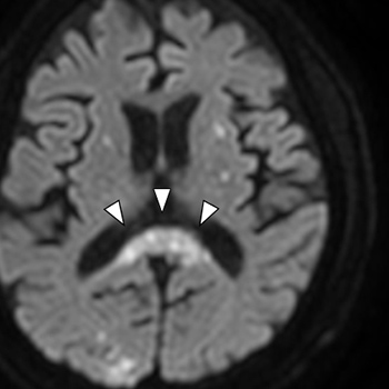
Background: Mild encephalitis/encephalopathy and a reversible splenial lesion (MERS) is a clinicoradiological syndrome with an unknown pathogenic mechanism, which usually involves children. Thus, adult-onset MERS is quite rare.
Case presentation: A 71-year-old man, undergoing haemodialysis due to diabetes-induced chronic kidney disease, manifested a persistent fever and disorientation. Blood culture detected methicillin-resistant Staphylococcus aureus (MRSA), while echocardiography revealed vegetation in the aortic and mitral valves. Magnetic resonance imaging of the head revealed a fluid-attenuated inversion recovery-high, diffusion-weighted image-high lesion in the splenium of the corpus callosum, with a number of emboli. Accordingly, the patient was diagnosed with MERS induced by MRSA endocarditis.
Discussion: Neurological impairment by MERS can be reversible. However, the differential diagnosis of the disease includes ischaemic lesions, multiple sclerosis, malignant lymphoma, acute disseminated encephalomyelitis, and posterior reversible encephalopathy. Clinicians should consider these diseases when MERS is suspected.
|
Views: 702
HTML: 78
PDF: 309
|
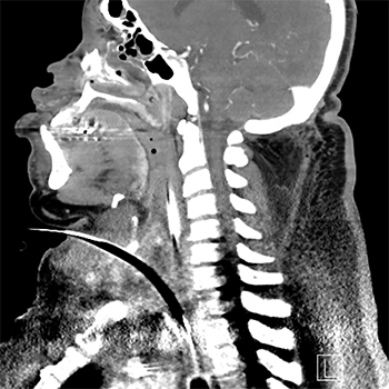
Although allergies to antibiotics are commonly stated, anaphylactic reactions are uncommonly reported. This is especially the case with reactions to fluoroquinolone antibiotics. Furthermore, airway emergencies are rare. We present a case of ciprofloxacin-induced acute airway obstruction and anaphylaxis, necessitating emergent surgical cricothyrotomy following respiratory distress.
|
Views: 667
HTML: 77
PDF: 561
|
SARS-CoV-2 has infected millions of people worldwide. Its cardiac presentations include myocarditis, arrhythmias and structural heart changes even in young and healthy individuals. The long-term sequelae of these manifestations are unknown. We describe a unique combination of complete heart block and atrial flutter in the setting of COVID-19. SARS-CoV-2 virulence mechanisms can cause fibrosis in the myocardium resulting in loss of sinus node dominance. The paradoxical finding of atrial flutter and complete heart block is very rare. Prompt cardiac evaluation and electrophysiological testing are important. Cardiac magnetic resonance imaging (cMRI) and endomyocardial biopsies are the gold standard investigations. Anticoagulation should be administered until atrioventricular synchrony is achieved.

|
Views: 759
HTML: 83
PDF: 308
|
An 80-year-old patient was admitted to the internal medicine department for binocular diplopia and hearing loss with sudden onset. The patient had presented with SARS-CoV-2 infection 3 weeks previously and had been admitted to hospital. Complete work-up including autoimmunity, serum and LCR viral serology and MRI did not allow a diagnosis to be established. The hypothesis of a microvascular origin or the previous SARS-CoV-2 infection was considered. The latter was retained in light of the temporal relationship, the absence of other pathologies after exhaustive work-up, and the clinical evolution.
|
Views: 553
HTML: 451
PDF: 394
|
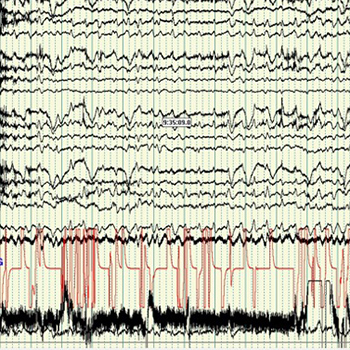
Hepatic encephalopathy is a common complication in chronic liver disease and cirrhosis. Here we describe two patients with hepatic encephalopathy who did not respond to standard empiric treatment and were found to have non-convulsive status epilepticus. Both patients improved with antiepileptic therapy. Non-convulsive status epilepticus should be considered in the differential diagnosis of patients with suspected hepatic encephalopathy who do not respond to empiric treatment.

|
Views: 824
HTML: 1709
PDF: 540
|
Giant cell arteritis (GCA) and polymyalgia rheumatica (PMR) are both rheumatological diseases of the elderly with a strong association with each other and which rarely present with normal inflammatory markers. Here we present the case of a 61-year-old Caucasian woman who had typical symptoms of both diseases. At the time of presentation, her blood work showed normal inflammatory markers, but because of the high clinical suspicion for GCA, a temporal artery biopsy was done which was positive for giant cells and disruption of the internal elastic lamina. Our patient responded very well to treatment with oral steroids and steroid-sparing medication and was able to return to her normal life without experiencing any complications of the disease. By sharing our case, we aim to increase the awareness of medical personnel regarding the importance of focusing on the clinical presentation as well as the laboratory and pathological aspects of diagnosing GCA and PMR.
|
Views: 565
HTML: 348
PDF: 281
|
Complete blood counts are frequently collected from cancer patients, but laboratory findings may be misleading. Secondary polycythemia can occur in renal cell carcinoma (RCC) due to erythropoietin (EPO) stimulation. Therefore, complete blood counts should be closely monitored to prevent complications such as thrombosis. We discuss the case of a 47-year-old man with metastatic RCC who presented with secondary polycythemia that improved with chemotherapy. His secondary erythrocytosis was anticipated, but his haemoglobin levels were lower than expected after therapy. This article discusses the treatment and diagnosis of secondary polycythemia in patients with RCC.
|
Views: 383
HTML: 57
PDF: 411
|
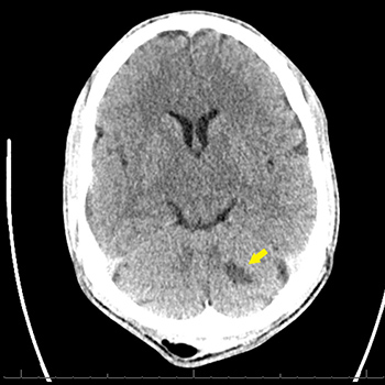
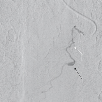
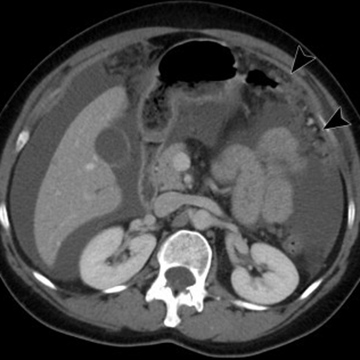
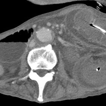
We report a case of a bronchial artery pseudoaneurysm presenting as acute retrosternal pain. We want to discuss and to announce the extremely rare finding of a bronchial artery pseudoaneurysm. Bronchial artery aneurysms and pseudoaneurysms are uncommon; however, missing this diagnosis is associated with significant morbidity and mortality. When suspecting this pathology urgent CT angiography and selective angiography (DSA) are crucial. Urgent treatment with transarterial embolization is preferred.
|
Views: 473
HTML: 165
PDF: 279
|
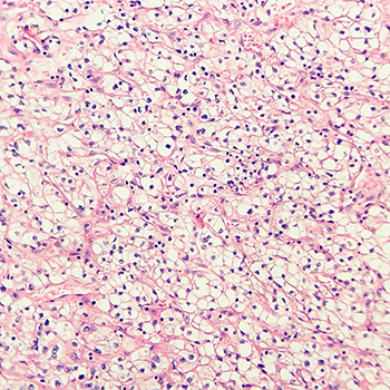
A myeloid sarcoma is an extramedullary tumour arising from infiltration by leukemic cells at an anatomic site other than the bone marrow. Most commonly it precedes acute myeloid leukaemia but occasionally occurs simultaneously. It may also be associated with myeloproliferative neoplasms, myelodysplastic syndrome and the blast phase of chronic myeloid leukaemia.
The most common sites for extramedullary tumours are bone, periosteum, soft tissue, lymph node and skin. Although this disease can affect a wide range of body sites, there are very few reports of peritoneal myeloid sarcoma or cavity effusion.
The authors present the case of a 68-year-old man with myelodysplasia-related acute myeloid leukaemia and peritoneal myeloid sarcoma with myeloid ascites. The definitive diagnosis is challenging, requires a high level of suspicion, and relies on the exclusion of all alternative diagnoses and especially on complementary tests such as flow cytometry and immunohistochemistry analysis of ascitic fluid in order to detect the immature myeloid cells.
|
Views: 323
HTML: 50
PDF: 191
|
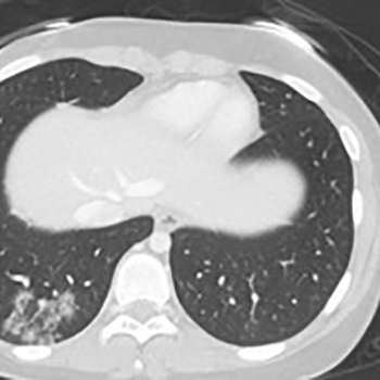
Hydropneumopericardium is a rare event with a risk of serious complications. Timely diagnosis and treatment is important as it can improve prognosis. We report the case of a 77-year-old male patient who presented with acute interscapular pain which developed during a meal. An oesophago-pericardial fistula was found in the context of malignant oesophageal disease.

|
Views: 605
HTML: 296
PDF: 424
|
We report the case of a 61-year-old man admitted to our emergency department with fever. At admission, he was hypotensive and tachycardic. In the initial investigation, elevation of inflammatory parameters, acute kidney injury (Kidney Disease Improving Global Outcomes (KDIGO) 3), hyperbilirubinemia, and hepatic cytocholestasis were evident. Empirical antibiotic therapy was started, after sepsis was assumed without an identifiable cause. His condition took an unfavorable clinical course, with respiratory failure, hepatosplenomegaly, pancytopenia, hyperferritinemia and hypofibrinogenemia. Microbial culture studies and a general immunological study were negative and lymphoproliferative disease was therefore excluded. Bone marrow aspirate revealed hemophagocytosis without granulomas. A diagnosis of hemophagocytic lymphohistiocytosis was assumed and pulse methylprednisolone therapy initiated. As this resulted in only a transient improvement, immunoglobulin and rituximab were initiated as a second-line therapy. The patient sadly had an unfavorable outcome despite all measures undertaken. In the postmortem study, Mycobacterium tuberculosis complex was isolated in the bone marrow aspirate, which led to the postmortem diagnosis of disseminated tuberculosis and angioinvasive pulmonary aspergillosis. The clinical presentation of disseminated tuberculosis is non-specific and hemophagocytic lymphohistiocytosis is one of its rare presentations. The mortality rate of hemophagocytic lymphohistiocytosis is high and increases with delayed diagnosis of the underlying condition and respective treatment.
|
Views: 882
HTML: 136
PDF: 383
|
Introduction: The detection of pneumococcal antigens in urine is an alternative to gram staining, and their culture is central to the diagnosis of pneumococcal pneumonia. We present a case of the false-positive detection of urinary Streptococcus species pneumococcal antigen with a BinaxNOW test. This resulted in delayed diagnosis of a liver abscess.
Case description: A 70-year-old woman presented to the emergency department with a 1-day history of chills and difficulty walking. She had a fever and her physical examination was normal. Non-contrast chest computed tomography (CT) revealed a slight ground-glass opacity in the left lower lobe. Laboratory tests revealed liver injury and elevated C-reactive protein levels. A urinary pneumococcal antigen test was positive, and she was diagnosed with acute bronchopneumonia caused by Streptococcus pneumoniae. She was treated with ceftriaxone. However, abdominal contrast-enhanced CT performed the next day revealed portal vein thrombus and a left lobe liver abscess. Streptococcus constellatus was detected in a puncture specimen of the liver abscess. It was concluded that the positive urinary pneumococcal antigen test was a false-positive owing to Streptococcus infection.
Discussion: False-positive results might be explained by the presence of C-polysaccharide antigens in the cell wall of S. pneumoniae. The positive urinary antigen test together with the finding of slight ground-glass opacity in the left lung on chest CT initially led to misdiagnosis. False positives may result in misdiagnosis and unnecessary antimicrobial therapy.
Conclusion: The overuse of the pneumococcal urinary antigen tests can lead to false positives and misdiagnosis.

|
Views: 876
HTML: 152
PDF: 350
|
Introduction: Lymphoplasmacytic lymphoma (LPL) is a rare low-grade B-cell neoplasm that accounts for approximately 2% of all haematological malignancies. Most patients have the clinical syndrome of Waldenstrom macroglobulinemia (WM), which is defined as LPL with an associated immunoglobulin M (IgM) serum monoclonal protein. Roughly 5% of LPL patients secrete non-IgM paraproteins (e.g., IgG, IgA, kappa, lambda) or are non-secretory.
Case description: We report the case of a 41-year-old woman who was diagnosed with non-IgM LPL with lambda light chain monoclonal paraprotein production and normal serum immunoglobulin levels. The MYD88 L265P mutation was detected on fluorescence in-situ hybridization (FISH) analysis of the bone marrow. The patient underwent treatment with a combination of ibrutinib and rituximab. There was an initial response but she died 8 months after diagnosis.
Discussion: Non-IgM LPL poses diagnostic and therapeutic challenges to clinicians as it is an exceptionally rare malignancy with a heterogeneous clinicopathological presentation and scarce literature. Among non-IgM LPL cases, those with lambda light chain production are even more rare. To the best of our knowledge, none have been reported to date. The addition of MYD88 L265P testing to the diagnostic armamentarium of non-IgM LPL cases is advisable for potential therapeutic reasons.
|
Views: 595
HTML: 90
PDF: 287
|
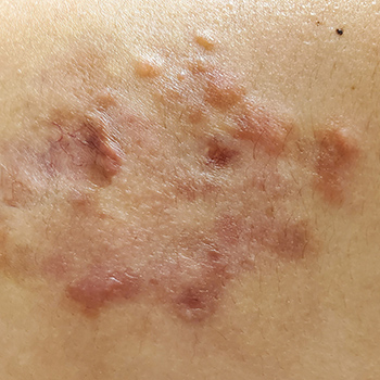
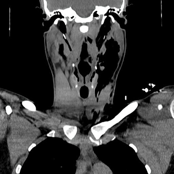
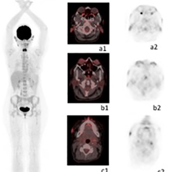
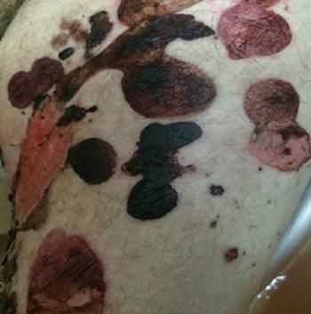
Rosai–Dorfman disease (RDD) is a rare disorder characterized by the proliferation of histiocytes in lymph nodes. It can occur anywhere in the body but commonly involves the cervical area of the neck. Its clinical presentation varies and patients with skin manifestations may develop papules, nodules, plaques, or pustules. Histologically, it typically presents with emperipolesis, where intact lymphocytes are found within histiocytes. The definitive treatment of RDD is not well established given the rarity of the disease and indeed skin lesions can regress spontaneously. Therapeutic treatment options include cryotherapy, radiation, or topical agents such as steroids or retinoids. Here we describe the case of a 24-year-old Hispanic female who presented with skin manifestations which proved to be histologically positive for Rosai–Dorfman disease. The patient clinically improved following the administration of intralesional steroids.
|
Views: 325
HTML: 42
PDF: 279
|
A 79-year-old man was admitted to our hospital due to pleural empyema. After 4 weeks of antimicrobial therapy and pleural drainage, he recovered but complained of new-onset abdominal pain. Abdominal computed tomography revealed adhesive small bowel obstruction and a nasointestinal ileus tube with intermittent suction was inserted. This procedure initially decreased his abdominal pain, but severe abdominal pain and vomiting developed 3 days later. Repeat abdominal computed tomography revealed jejuno-jejunal intussusception due to the nasointestinal ileus tube. Our patient was initially treated conservatively. However, he underwent surgical reduction due to clinical deterioration 1 day after diagnosis and died from a surgical complication 19 days later. Intussusception is a rare but fatal complication caused by placement of a nasointestinal ileus tube in the small intestine. Because urgent operative reduction is needed to avoid intestinal resection in most cases, early diagnosis and surgical reduction of intussusception are critical.
|
Views: 425
HTML: 133
PDF: 253
|
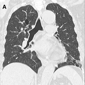
Neuromyelitis optica spectrum disorder (NMOSD) is associated with other autoimmune disorders and probably with cryptogenic organizing pneumonia (COP) as well. Here we present the case of a 14-year-old girl presenting with typical NMOSD together with radiological evidence of COP. Our case is unique as the previous two reports of this association were in elderly patients.
|
Views: 515
HTML: 105
PDF: 508
|
Subcutaneous emphysema is the presence of air beneath the skin’s soft tissues. It can result from medical conditions, trauma or iatrogenic causes. The occurrence of subcutaneous emphysema after a dental procedure is rare. Although it is mostly a benign and self-limiting complication, the consequences may be severe and life-threatening. We report the case of a 20-year-old man who presented to the emergency department with swelling of his face and neck after dental treatment. The diagnosis of subcutaneous emphysema and pneumomediastinum was made based on physical examination and a computerized tomography scan.
|
Views: 631
HTML: 108
PDF: 361
|
Rosai-Dorfman-Destombes disease (RDD) or sinus histiocytosis with massive lymphadenopathy is a rare non-Langerhans cell histiocytosis of unknown cause. The disease often manifests as bilateral painless cervical lymphadenopathy associated with systemic symptoms such as fever and weight loss. Extranodal disease is also frequent and can involve any organ, mostly skin, nasal cavity, bone, and retro-orbital tissue. Swelling of cartilaginous tissues such as ear helix or laryngeal structures may mimic the entity known as relapsing polychondritis. Although spontaneous remission is the most expected evolution, some cases require systemic treatment with prednisone, methotrexate or cytotoxic agents, with variable success rates. In this respect, since somatic variants in genes involved in the mitogen-activated protein kinase (MAPK) pathway have been observed to play a pathogenic role in RDD, the use of therapies targeting these pathogenic variants seems to be a reasonable strategy. Here we present a case of RDD with extensive extranodal involvement that showed a rapid and complete response to the MEK inhibitor cobimetinib
|
Views: 1373
HTML: 285
PDF: 793
|
It is increasingly recognized that SARS-CoV-2 infection and COVID-19 vaccines have been associated with skin disorders, including pityriasis rosea. It has been reported that pityriasis rosea has been triggered by several vaccines, as a rare side-effect. We present two cases of COVID-19 vaccine-induced pityriasis rosea. Skin lesions appeared in a 49-year-old female 8 days after the first dose of the BNT162b2 mRNA vaccine and in a 53-year-old male 7 days after the second dose of the same vaccine. The exanthem was self-limited in both patients over a period of a month.

|
Views: 372
HTML: 79
PDF: 300
|
Yersinia enterocolitica infection is an uncommon but potentially fatal zoonosis, especially when it culminates with septicaemia. Post-infectious complications like reactive arthritis and erythema nodosum are well described in the literature. On the other hand, the association between yersinosis and autoimmune haemolytic anaemia (AIHA) has only been established in one clinical case. We present a case of an 87-year-old man admitted for AIHA, whose was successfully treated only after the identification and treatment of yersinosis.
|
Views: 830
HTML: 67
PDF: 476
|
Bullous pemphigoid is a rare autoimmune dermatologic disease that usually occurs in the elderly. Mucous membrane lesions occur in about 10–35% of patients and are almost always limited to the oral mucous membrane. Esophageal involvement is very rare (4% of cases) and usually presents with chest pain, dysphagia, and odynophagia, though patients are frequently asymptomatic. We report the case of newly diagnosed bullous pemphigoid in a 76-year-old man with a past medical history of dementia. He presented with cutaneous manifestations but also severe gastrointestinal bleeding due to extensive esophageal involvement. Although bullous pemphigoid is mainly a skin disease, mucous membrane lesions should not be overlooked as they are associated with an even poorer outcome. A high index of suspicion for esophageal involvement is needed as its presentation can be fatal, as with our patient.
| 2.1 = | 1.730 Cit. to date |
| 842 Docs. to date |


Publisher
Official Journal of the
European Federation of Internal Medicine
www.efim.org
Publisher: SMC media Srl
Via Giovenale, 7 - 20136 Milan - Italy
P.IVA 07626490960
info@ejcrim.com
www.ejcrim.com - ISSN: 2284-2594 - © EFIM 2014-2023, Published by SMC Media srl, Italy - Privacy policy

