A 28-Year-Old Woman with Ascites and Multiple Focal Spleen Lesions
  • Alice Pirovano
    Department of Translational Medicine, Università del Piemonte Orientale, Novara, Italy
  • Erica Matino
    Department of Translational Medicine, Università del Piemonte Orientale, Novara, Italy
  • Erika Zecca
    Department of Translational Medicine, Università del Piemonte Orientale, Novara, Italy
  • Martina Costanzo
    Department of Translational Medicine, Università del Piemonte Orientale, Novara, Italy
  • Alessandro Croce
    Department of Translational Medicine, Università del Piemonte Orientale, Novara, Italy
  • Monica Leutner
    Azienda Ospedaliero-Universitaria Maggiore della Carità, Novara, Italy
  • Raffaele Romito
    Azienda Ospedaliero-Universitaria Maggiore della Carità, Novara, Italy
  • Mario Pirisi
    Department of Translational Medicine, Università del Piemonte Orientale, Novara, Italy

Keywords

Ascites, splenomegaly, lymphoma, disseminated intravascular coagulation, diagnostic splenectomy

Abstract

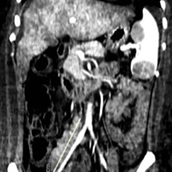
Serous effusions complicating the course of lymphomas occur commonly in the pleural space but seldom in the peritoneum, where they most often present as chylous ascites with diagnostic cytology. Almost invariably, in these rare cases, the serum to ascites albumin gradient is low. We describe a 28-year-old woman with anasarca, ascites and a serum to ascites albumin gradient of 1.1 g/dl, consistent with portal hypertension. No tumour cells were detected in the ascitic fluid. However, a CT scan of the chest and abdomen disclosed liver and spleen enlargement and multiple enlarged retroperitoneal lymph nodes, suspicious for a lymphoproliferative disorder. Bone marrow aspiration and biopsy were not diagnostic, so a decision was made to proceed with a splenectomy despite the onset of low-grade disseminated intravascular coagulation.
Surgery was uneventful. Diffuse large B cell lymphoma was diagnosed. A liver biopsy taken at the time of surgery demonstrated that the liver parenchyma was massively infiltrated by reactive T lymphocytes surrounding rare large CD20+ tumour cells. This infiltrate had likely led to increased portal pressure attended by ascites formation, which resolved completely after chemotherapy. The case emphasizes the rewards of pursuing a diagnosis supported by a high prior probability even in the presence of apparently discordant laboratory findings, as well as the importance of performing a diagnostic splenectomy in case of splenomegaly with unexplained focal lesions.

VIEW THE ENTIRE ARTICLE

References

  • Runyon BA. Management of adult patients with ascites due to cirrhosis. Hepatology 2004;39:841–856.

  • Riquelme A, Calvo M, Salech F, Valderrama S, Pattillo A, Arellano M, et al. Value of adenosine deaminase (ADA) in ascitic fluid for the diagnosis of tuberculous peritonitis: a meta-analysis. J Clin Gastroenterol 2006;40:705–710.

  • Schouten JN, Garcia-Pagan JC, Valla DC, Janssen HL. Idiopathic noncirrhotic portal hypertension. Hepatology 2011;54:1071–1081.

  • Das DK. Serous effusions in malignant lymphomas: a review. Diagn Cytopathol 2006;34:335–347.

  • Almakdisi T, Massoud S, Makdisi G. Lymphomas and chylous ascites: review of the literature. Oncologist 2005;10:632–635.

  • O’Reilly RA. Splenomegaly in 2,505 patients at a large university medical center from 1913 to 1995. 1963 to 1995: 449 patients. West J Med 1998;169:88–97.

  • Caremani M, Occhini U, Caremani A, Tacconi D, Lapini L, Accorsi A, et al. Focal splenic lesions: US findings. J Ultrasound 2013;16(2):65–74.

  • Thipphavong S, Duigenan S, Schindera ST, Gee MS, Philips S. Nonneoplastic, benign, and malignant splenic diseases: cross-sectional imaging findings and rare disease entities. AJR Am J Roentgenol 2014;203:315–322.

  • Pugalenthi A, Bradley C, Gonen M, Do KG, Strong V, Jarnagin W, et al. Splenectomy to treat splenic lesions: an analysis of 148 cases at a cancer center. J Surg Oncol2013;108:521–525.

  • Huang CJ, Pitt HA, Lipsett PA, Osterman FA Jr, Lillemoe KD, Cameron JL, et al. Pyogenic hepatic abscess. Changing trends over 42 years. Ann Surg 1996;223:600–607.

  • Lee WK, Van Tonder F, Tartaglia CJ, Dagia C, Cazzato RL, Duddalwar VA, et al. CT appearances of abdominal tuberculosis. Clin Radiol 2012;67:596–604.

  • World Health Organization. Global tuberculosis report 2018. Available from https://www.who.int/tb/publications/global_report/en/ (accessed 17 April 2019).

  • Compérat E, Bardier-Dupas A, Camparo P, Capron F, Charlotte F. Splenic metastases: clinicopathologic presentation, differential diagnosis, and pathogenesis. Arch Pathol Lab Med 2007;131:965–969.

  • Saboo SS, Krajewski KM, O'Regan KN, Giardino A, Brown JR, Ramaiya N, et al. Spleen in haematological malignancies: spectrum of imaging findings. Br J Radiol 2012;85:81–92.

  • Olson MC, Atwell TD, Harmsen WS, Konrad A, King RL, Lin Y, et al. Safety and accuracy of percutaneous image-guided core biopsy of the spleen. AJR Am J Roentgenol 2016;206:655–659.

  • McInnes MD, Kielar AZ, Macdonald DB. Percutaneous image-guided biopsy of the spleen: systematic review and meta-analysis of the complication rate and diagnostic accuracy. Radiology 2011;260:699–708.

  • Weledij EP. Benefits and risks of splenectomy. Int J Surg 2014;12:113–119.

  • Glatstein E, Guernsey JM, Rosenberg SA, Kaplan H. The value of laparotomy and splenectomy in the staging of Hodgkin's disease. Cancer 1969;24709–718.

  • Veronesi U, Musumeci R, Pizzetti F, Gennari L, Bonadonna G. Proceedings: The value of staging laparotomy in non-Hodgkin's lymphomas (with emphasis on the histiocytic type). Cancer 1974;33:446–459.

  • Matharoo GS, Afthinos JN, Gibbs KE. Trends in splenectomy: where does laparoscopy stand? JSLS 2014;18(4). pii: e2014.00239.

  • Chi S, Ikezoe T. Disseminated intravascular coagulation in non-Hodgkin lymphoma. Int J Hematol 2015;102(4):413–419.

  • Feugier P, Van Hoof A, Sebban C, Solal-Celigny P, Bouabdallah R, Fermé C, et al. Long-term results of the R-CHOP study in the treatment of elderly patients with diffuse large B-cell lymphoma: a study by the Groupe d'Etude des Lymphomes de l'Adulte. J Clin Oncol 2005;23(18):4117–4126.
  • Views: 1619
    HTML downloads: 499
    PDF downloads: 815


    Published: 2019-05-06
    Issue: Vol 6 No 5 (view)


    How to cite:
    1.
    Pirovano A, Matino E, Zecca E, Costanzo M, Croce A, Leutner M, Romito R, Pirisi M. A 28-Year-Old Woman with Ascites and Multiple Focal Spleen Lesions. EJCRIM 2019;6 doi:10.12890/2019_001061.