Atypical Kikuchi-Fujimoto disease: FDG-PET contribution to the diagnosis
  • Mutsuka Kurihara
    Department of General Medicine, Chiba University Hospital, Chiba, Japan
  • Yasutaka Yanagita
    Department of General Medicine, Chiba University Hospital, Chiba, Japan
  • Daiki Yokokawa
    Department of General Medicine, Chiba University Hospital, Chiba, Japan
  • Yu Li
    Department of General Medicine, Chiba University Hospital, Chiba, Japan
  • Masatomi Ikusaka
    Department of General Medicine, Chiba University Hospital, Chiba, Japan

Keywords

Type A aortic dissection, Bentall procedure, atypical symptoms

Abstract

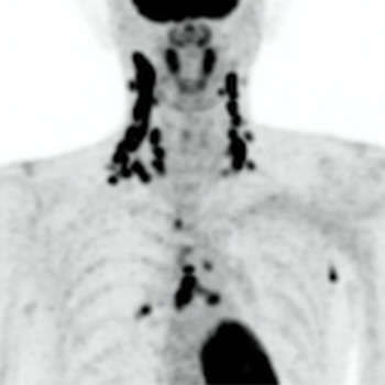
Kikuchi-Fujimoto disease (KFD), also called histiocytic necrotizing lymphadenitis, is more common in young women and typically presents with small, painful, localized cervical lymphadenopathy that resolves spontaneously within a few weeks. Laboratory findings are variable. As many as 40% of KFD cases are reported to be painless, and up to 22% to be generalized lymphadenopathy. Therefore, malignant lymphoma could be a differential diagnosis of KFD. A histopathologic diagnosis is needed when it is difficult to distinguish KFD from lymphoma. KFD typically shows small, highly accumulated cervical lymph nodes on fluorodeoxyglucose positron emission tomography (FDG-PET). This contrasts with malignant lymphoma, which tends to be associated with massive lymphadenopathy. In our case, a 40-year-old Japanese male presented with painless lumps in the right neck, accompanied by fever, night sweats, and loss of appetite. His symptoms and laboratory results worsened over a month. FDG-PET revealed highly accumulated uptake in cervical, mediastinal, and axillary lymph nodes. The PET imaging showed a small, high FDG uptake and contributed to the correct diagnosis of KFD. This case report highlights the importance of FDG-PET, which is a valuable diagnostic tool for KFD as it typically differentiates large clusters of small lymph nodes typical of KFD from normal lymph nodes.

VIEW THE ENTIRE ARTICLE

References

  • Perry AM, Choi SM. Kikuchi-Fujimoto Disease: A Review. Arch Pathol Lab Med 2018;142:1341-1346.
  • Kikuchi M. Lymphadenitis showing focal reticulum cell hyperplasia with nuclear debris and phagocytosis. Nippon Ketsueki Gakkai Zasshi 1972;35:378–380.
  • Fujimoto Y, Kozima Y, Hamaguchi K. Cervical necrotizing lymphadenitis: a new clinicopathological agent. Naika 1972;20:920–927.
  • Song JY, Lee J, Park DW, Sohn JW, Suh SI, Kim IS, et al. Clinical outcome and predictive factors of recurrence among patients with Kikuchi’s disease. Int J Infect Dis 2009;13:322-326.
  • Supari D, Ananthamurthy A. Kikuchi-Fujimoto disease: a study of 24 cases. Indian J Otolaryngol Head Neck Surg 2014;66:69–73.
  • Wang S, Du B, Li X, Li Y. Positron emission tomography/computed tomography hypermetabolism of Kikuchi-Fujimoto disease mimicking malignant lymphoma: a case report and literature review. J Int Med Res 2021;49:3000605211032859.
  • Views: 208
    HTML downloads: 34
    PDF downloads: 144


    Published: 2024-01-25
    Issue: 2024: Vol 11 No 2 (view)


    How to cite:
    1.
    Kurihara M, Yanagita Y, Yokokawa D, Li Y, Ikusaka M. Atypical Kikuchi-Fujimoto disease: FDG-PET contribution to the diagnosis. EJCRIM 2024;11 doi:10.12890/2024_004258.

    Most read articles by the same author(s)