EJCRIM 2023 CiteScore
| 2.1 = | 1.762 Cit. to date |
| 842 Docs. to date |
Last updated on 05 May, 2024
Updated monthly
Updated monthly
Powered by

|
Views: 1150
HTML: 690
PDF: 480
|
Non-traumatic haematomyelia is a rare finding of acute onset, which in most cases is the result of arteriovenous malformations (AVM), tumours, coagulation disorders or autoimmune conditions, but may also be secondary to treatment with anticoagulants and radiotherapy. We present the case of a 58-year-old woman with sudden onset cervical pain, followed by asymmetric diminution of strength in the upper limbs with reduced pain sensitivity. The diagnosis of AVM at the C7 and D1 levels was made following cervico-dorsal magnetic resonance imaging and angiography. Treatment was embolization with immediate isolation of the AVM.
|
Views: 1301
HTML: 1420
PDF: 541
|
We describe a case of emphysematous pancreatitis, a rare and serious complication of acute pancreatitis, which has a high mortality rate.
|
Views: 1105
HTML: 143
PDF: 484
|
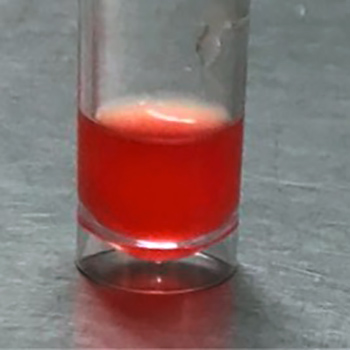
Few reports have been published on the correlation between plasma concentrations of rivaroxaban and clinical outcome in patients who have experienced venous thromboembolism. This article describes the case of a 44-year-old woman who experienced deep vein thrombosis during anticoagulation therapy with rivaroxaban, with evidence of repeated low plasma levels of the drug. We postulate that the determination of plasma rivaroxaban anti-Xa activity can be useful in the evaluation of anticoagulation therapy in selected cases.
|
Views: 1062
HTML: 485
PDF: 511
|
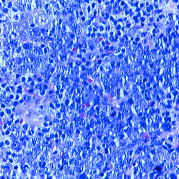
A 65-year-old man presented with a 2-year history of severe bilateral proptosis, palpable lymphadenopathy and moderate hepatosplenomegaly. A blood test was positive for hepatitis C infection. CT showed palpebral infiltrative lesions with regional progression through the temporal and masticatory spaces to the pharynx and hypopharynx causing almost complete airway obstruction. A palpebral biopsy was consistent with low-grade Bcl-2+ extra-nodal MALT non-Hodgkin B-cell lymphoma. The patient received six cycles of rituximab-based chemotherapy with clinical remission at 9-month follow-up. Bilateral proptosis is a rare presentation of several diseases. When brain CT excludes cavernous sinus pathology, thyroid ophthalmopathy or haematological malignancy should be considered.
|
Views: 1594
HTML: 489
PDF: 697
|
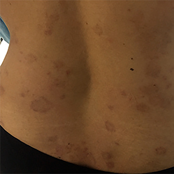
A 36-year-old woman with eosinophilic granulomatosis with polyangiitis (EGPA) presented with necrotic skin lesions and pulmonary infiltrates. There was eosinophilic vasculitis on skin biopsy, and substantial tissue eosinophilia in her bone marrow. She had unexplained worsening thrombocytopenia, which prompted a thrombophilia work-up. However, abnormalities in liver enzymes led to the extraordinary finding of portal vein thrombosis. Thrombocytopenia resolved with treatment with low molecular weight heparin. This case highlights the risk of hypercoagulability in eosinophilia specifically, and in EGPA. We suggest that thrombosis should be ruled out in all cases of EGPA.
|
Views: 896
HTML: 142
PDF: 466
|
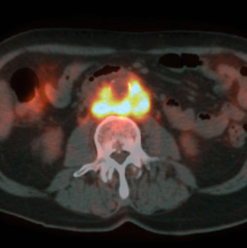
Aortitis is a rare condition and easily overlooked. It is defined as infectious or non-infectious inflammation of the aortic wall. This report describes two cases of aortitis, demonstrating the diagnostic difficulty and how diagnostic delay could have been reduced if early radiology had been performed. Due to the nature of aortitis, patient outcome can be improved considerably by timely diagnosis and treatment.
|
Views: 1095
HTML: 630
PDF: 558
|
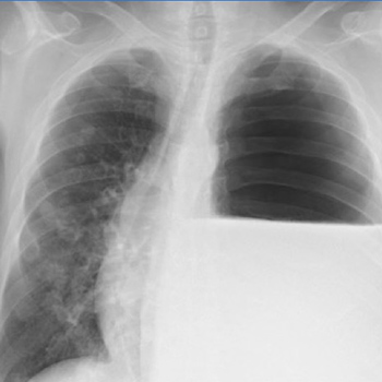
Although the current medical literature is limited, hydropneumothorax was described as far back as the 5th century BC. It is characterized by the presence of air and fluid in the pleural cavity and is an infrequent finding. Causes include trauma, iatrogenesis following thoracentesis, the presence of gas-forming organisms, tuberculosis and malignancy. Diagnosis is based on clinical and radiological features. We report a case of hydropneumothorax and present radiological images showing the distinctive features of this entity.
|
Views: 4075
HTML: 446
PDF: 938
|
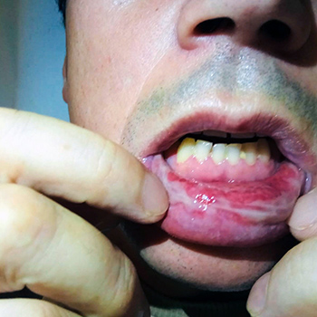
Mycoplasma pneumoniae (MP) is a common cause of respiratory infections and can be associated with extrapulmonary complications. MP mucositis has recently been described as a distinct endemic clinical entity called Mycoplasma pneumoniae-induced rash and mucositis (MIRM). The authors present the case of a 46-year-old man with atypical pneumonia associated with exuberant mucositis, conjunctival hyperaemia and positive serological assays for MP IgM. The patient was treated with azithromycin and systemic corticosteroid therapy. Supportive care including pain management, intravenous hydration and mucosal care was also given. There was complete resolution of the pneumonia and mucositis. The presence of atypical pneumonia with mucosal involvement without cutaneous lesions and a favourable clinical evolution led to the diagnosis of MIRM.
| 2.1 = | 1.762 Cit. to date |
| 842 Docs. to date |


Publisher
Official Journal of the
European Federation of Internal Medicine
www.efim.org
Publisher: SMC media Srl
Via Giovenale, 7 - 20136 Milan - Italy
P.IVA 07626490960
info@ejcrim.com
www.ejcrim.com - ISSN: 2284-2594 - © EFIM 2014-2024, Published by SMC Media srl, Italy - Privacy policy

