EJCRIM 2023 CiteScore
| 2.1 = | 1.751 Cit. to date |
| 842 Docs. to date |
Last updated on 05 April, 2024
Updated monthly
Updated monthly
Powered by


|
Views: 36
HTML: 2
PDF: 17
|
Euglycemic diabetic ketoacidosis (euDKA) is a rare but severe metabolic complication of diabetes mellitus characterised by elevated anion gap metabolic acidosis despite normal or mildly elevated blood glucose levels. Sodium-glucose cotransporter 2 inhibitors (SGLT2i) have emerged as effective antidiabetic medications, yet their use is associated with an increased risk of euDKA, especially when coupled with insulin dose reduction.
We present the case of a 50-year-old male with a 20-year history of diabetes mellitus, initially managed with insulin and metformin, who developed euDKA following the introduction of empagliflozin and sitagliptin alongside a reduction in insulin therapy. Despite normoglycaemia the patient exhibited symptoms of ketoacidosis, including chronic fatigue, polydipsia, and polyuria.
Diagnostic workup revealed metabolic acidosis, elevated inflammatory markers, acute kidney injury and ketonuria. Subsequent specialised laboratory tests confirmed type 1 diabetes mellitus (T1DM) with the presence of anti-glutamic acid decarboxylase (anti-GAD) antibodies and the absence of C-peptide secretion. Management involved fluid therapy, intravenous insulin and glucose administration.
This case underscores the diagnostic challenges of euDKA and emphasises the importance of differentiating between T1DM and T2DM, as management strategies vary significantly. Patient education on insulin therapy and injection techniques is crucial to prevent complications such as improper insulin delivery and dose reduction, which can precipitate euDKA.
In conclusion, clinicians should be vigilant for euDKA in patients on SGLT2 inhibitors, particularly when insulin dose reduction is involved. Comprehensive patient education and accurate differentiation between diabetes types are essential for timely diagnosis and optimal management, thereby reducing the risk of severe complications.
|
Views: 173
HTML: 43
PDF: 109
|
Background: Community-acquired bacterial meningitis in adults represents one of the most severe infectious diseases worldwide with potentially life-threatening medical complications. Several infectious agents can cause acute meningitis. Although group B Streptococcus is more prevalent in newborns, infection can also lead to meningitis in older adults, particularly those with underlying health issues.
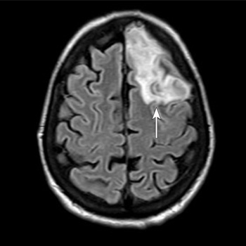
Case Description: A 53-year-old woman with a body mass index of 28.7 kg/m2, type 2 diabetes mellitus, and dyslipidaemia presented to the emergency department of Santa Maria della Stella Hospital (Orvieto, Italy) with confusion, low-grade fever, echolalia, and hyperglycaemia. Computed tomography scans of the brain revealed a hypodensity in the left anterior frontal lobe and an osteodural defect of the rhinobase. Meningitis was suspected and empiric broad-spectrum antibiotic therapy with corticosteroids and insulin were administered while the results of the cerebrospinal fluid analysis confirmed the diagnosis of group B Streptococcus meningitis. Repeat imaging at 48 hours revealed enlargement of the hypodense lesion. The frontal assessment battery indicated deficits in executive functions. Prompt treatment led to rapid clinical improvement. Following the restoration of euglycemic status and hemodynamic stabilization, a follow-up magnetic resonance imaging confirmed the ischaemic lesion and showed cerebrospinal fluid in the sella turcica. The patient was then transferred to neurorehabilitation.
Conclusions: The complex interactions among multiple risk factors resulted in an atypical clinical case of group B Streptococcus meningitis, which was promptly treated with empiric antibiotic therapy to mitigate neurocognitive deficits.
|
Views: 84
HTML: 8
PDF: 69
|
Introduction: Caustic substances ingestion results in a complex syndrome. The patient characteristics and severity of injury are important prognostic predictors. The monitoring of clinical changes and the multidisciplinary approach are necessary to prevent death in the early stages of the poisoning.
Case description: The case report describes the suicide of a woman by ingestion of a large amount of 15% sulfuric acid for suicidal purposes (15–20 ml). The initial conditions were stable, and no changes were found on a CT scan. However, the main sign was a severe metabolic acidosis. After 7 hours, haematemesis and oedema of the larynx appeared, and oro-tracheal intubation and ICU admission were necessary. Consequent progressive haemodynamic deterioration with persistent severe metabolic acidosis, increasing lactates and septic shock appeared. A new CT scan with contrast was performed 22 hours later detecting diffuse perforations and liquid in pleurae and abdomen. A pleural sample showed necrotic liquid. The death was 24 hours after ingestion and no surgical treatment was performed because of the diffuse lesions to the thoracoabdominal districts.
Conclusions: Early detection of gastroenteric lesions and the monitoring of clinical changes are mandatory to avoid the death of the patient. Gastroenteric perforations require an immediate radiological evaluation and surgical treatment. The clinical case report states the severity of prognosis was related to high doses of sulfuric acid ingestion. The immediate metabolic acidosis is related to quick subsequent severe systemic pathological lesions of the gastrointestinal tract. The severity of absorption metabolic acidosis, consequently, may be an early and prognostic sign of the late chest and abdominal lesions.
|
Views: 163
HTML: 9
PDF: 71
|
Mitral valve prolapse (MVP) is a primary valvular disease of the mitral valve with a prevalence of 2.4% of the general population. Valve abnormalities range from simple fibroelastic deficiency of the leaflets to diffuse myxomatous degenerative changes. MVP is a usually a benign condition. However, the scattered reports of sudden cardiac death in patients with MVP in the absence of severe mitral insufficiency or coronary artery disease suggest the existence of a malignant phenotype of MVP. We report a case of a young female who survived life-threatening arrhythmias and cardiac arrest and was found to have characteristic features of the malignant phenotype of MVP and had an implantable cardioverter defibrillator as a secondary prevention.
|
Views: 124
PDF: 88
HTML: 26
|
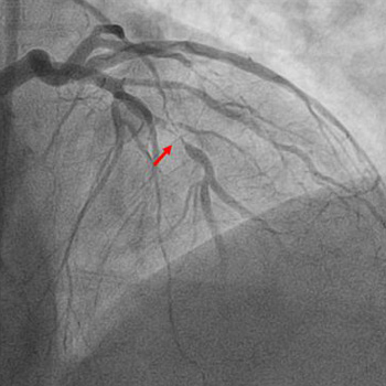
An acute ST-elevation myocardial infarction (STEMI) followed by reinfarction within a short period of time is typically due to stent thrombosis. However, a STEMI caused by occlusion of one vessel followed by a repeat infarction due to occlusion of a different vessel which was seemingly innocent a few hours earlier is extremely rare. We present the case of a 61-year-old male with a past medical history of prediabetes, hyperlipidemia, tobacco use, and gastroesophageal reflux disease who presented to the emergency department with complaints of chest pain. His initial electrocardiogram (EKG) revealed ST elevation in leads II, III and aVF with reciprocal changes in leads I and aVL. He promptly underwent cardiac catheterization and had percutaneous coronary intervention with placement of two drug-eluting stents (DES) in the right coronary artery (RCA). At that time coronary angiography revealed 50% stenosis of the left anterior descending (LAD) artery and 60% stenosis of the second diagonal branch artery. Shortly after the procedure he was asymptomatic, and the post procedure EKG demonstrated resolution of the ST elevations. However, within 2 hours he developed chest pain and was found to have new ST elevations in the anterolateral leads. Repeat cardiac catheterization revealed patent RCA stents with subtotal occlusion of the LAD and another DES was placed. After the second procedure the patient remained hemodynamically stable, EKG changes resolved, and he was kept on eptifibatide infusion for 18 hours after which he was switched to dual antiplatelet therapy and ultimately discharged home.

|
Views: 136
HTML: 7
PDF: 95
|
Introduction: Sudden onset of reduced consciousness, psychomotor agitation and mydriasis are all indicative of an anticholinergic toxidrome. It is important to note that numerous drugs, as well as certain herbs and plants, possess anticholinergic properties.
Case description: An 84-year-old female patient had sudden nocturnal onset of uncoordinated hand movements and altered mental status. Shortly after, the patient’s 83-year-old husband developed symptoms of dysarthria, gait ataxia, vertigo, and delirium.
Conclusion: Anticholinergic syndrome consists of a combination of central and peripheral anticholinergic symptoms. Physostigmine given intravenously resulted in rapid reversal of symptoms. Thorn apple seeds had been accidentally ingested and were identified as the cause.
|
Views: 263
HTML: 21
PDF: 150
|
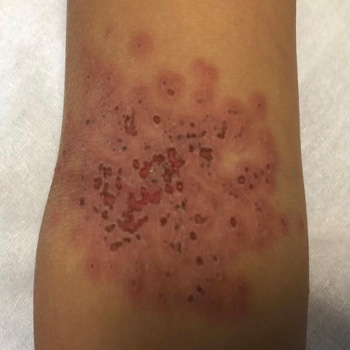
Introduction: Kaposi’s varicelliform eruption (KVE), also known as eczema herpeticum or eczema vaccinatum, is an acute dermatosis that affects patients with chronic dermatopathies. The diagnosis is primarily clinical and is characterised by the presence of a vesicular exanthema on physical examination. The exanthema subsequently evolves into crusted lesions with typical circular ulcerations in ‘punched-out’ areas on the skin affected by the underlying dermatopathy.
Case description: We present the case of a 6-year-old patient who presented to the Paediatric Emergency department with skin lesions consistent with eczema herpeticum. The patient’s management was initially outpatient; however, due to the slow progression of the condition, hospitalisation and intravenous antiviral treatment were initiated.
Discussion: KVE affects patients with chronic dermatoses, especially atopic dermatitis. It is important to know the clinical presentation for an early suspicion. KVE is a medical emergency that requires prompt diagnosis and treatment. It can progress to secondary viraemia, which can be fatal in up to 10% of immunocompetent individuals and up to 50% of immunocompromised individuals. It is important to be aware of this condition and to start early treatment with antivirals, especially given the high prevalence of atopic dermatitis in our population. This condition is one of the most serious complications that can occur in these patients.
|
Views: 153
HTML: 15
PDF: 125
|
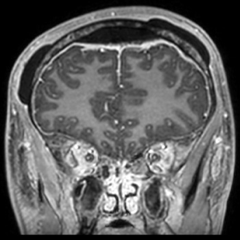
Introduction: Pott’s puffy tumour is a rare entity defined by the presence of a subperiosteal abscess of the frontal bone associated with frontal osteomyelitis. Several predisposing conditions can lead to this entity, such as frontal sinusitis.
Case description: We report the case of a 15-year-old patient who presented to the emergency department for headache, fever and forehead swelling. Computed tomography revealed severe pansinusitis complicated by a subperiosteal abscess associated with frontal osteomyelitis, leading to the diagnosis of Pott’s puffy tumour. The management combined intravenous antibiotics and surgical drainage of both the sinusitis and subperiosteal abscess.
Discussion: Pott’s puffy tumour represents a rare but serious complication of frontal sinusitis. Clinicians should be aware of this potential complication as the diagnosis can be challenging at an early stage but may influence the subsequent prognosis.
|
Views: 163
HTML: 9
PDF: 136
|
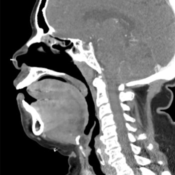
Spontaneous bleeding into the upper airways is a rare and potentially life-threatening complication of chronic anticoagulation. There are scarce cases in the literature demonstrating upper airway haematomas secondary to warfarin use, which is the predominant anticoagulant used by clinicians despite having a complex pharmacokinetic and pharmacodynamic profile. We report a compelling case featuring warfarin-induced sublingual haematoma, managed conservatively through the reversal of anticoagulation using fresh frozen plasma complemented by vigilant monitoring within the Intensive Care Unit (ICU).
|
Views: 223
HTML: 40
PDF: 152
|
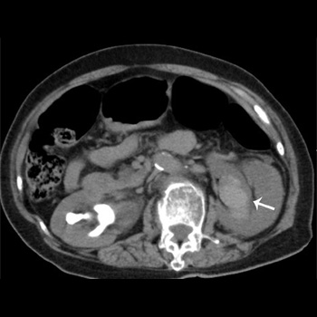
Subepithelial haemorrhage of the renal pelvis is a rare cause of haematuria and can be diagnosed based on radiographic findings. This haemorrhage often appears as a non-enhancing hyperdense mass in the renal pelvis on computed tomography, which sometimes results in unnecessary nephrectomy because it can mimic renal neoplasms. It can be managed conservatively, and its prognosis is generally benign. We report a case of renal pelvic haemorrhage complicating emphysematous pyelonephritis that needed emergent nephrectomy. Our case highlights the importance of careful observation for complications of urinary tract infection, although complications are rare.
| 2.1 = | 1.751 Cit. to date |
| 842 Docs. to date |


Publisher
Official Journal of the
European Federation of Internal Medicine
www.efim.org
Publisher: SMC media Srl
Via Giovenale, 7 - 20136 Milan - Italy
P.IVA 07626490960
info@ejcrim.com
www.ejcrim.com - ISSN: 2284-2594 - © EFIM 2014-2024, Published by SMC Media srl, Italy - Privacy policy

