
Keywords
Necrotizing fasciitis
Abstract
Objective: Necrotizing fasciitis is a difficult diagnosis with a very high mortality. However, thermal imaging has the potential to identify increasing skin temperature and rapid progression.
Materials and methods: We used repeat photographs taken with a thermal camera to visualize changes in skin temperature over time.
Results: An unstable male patient presented at the emergency department. Thermal imaging showed increased skin temperature of his left foot with a rapid increase and progression in extent within 1 hour. Necrotizing fasciitis was suspected and later confirmed.
Conclusions: We believe thermal imaging could be an important adjunct for the diagnosis of suspected necrotizing fasciitis.
References
Views: 1155
HTML downloads: 115
PDF downloads: 509
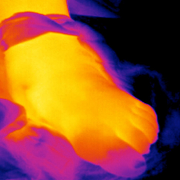
Figure 1a, conventional and thermographic images at arrival. At this time, the hottest area at the ankle was 35°C. downloads: 0
Figure 1b, conventional and thermographic images after 1 hour. At this time, the hottest area at the ankle was 37°C, showing a distal decrease and proximal increase in surface temperature. downloads: 0
Published:
2017-10-18
Issue:
Vol. 4 No. 10
(view)


