EJCRIM 2023 CiteScore
| 2.1 = | 1.762 Cit. to date |
| 842 Docs. to date |
Last updated on 05 May, 2024
Updated monthly
Updated monthly
Powered by

|
Views: 645
HTML: 74
PDF: 368
|
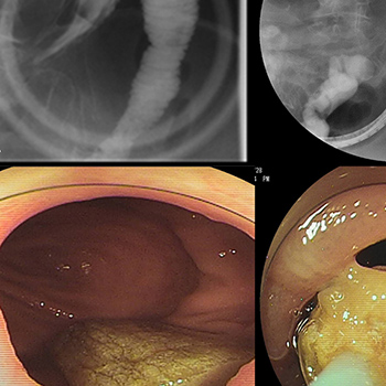
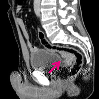
An enterolith in Crohn’s disease is an uncommon but serious condition because it can cause intestinal obstruction. Endoscopic treatment to remove the enterolith is attempted first, but is sometimes difficult owing to poor accessibility of the endoscope. In such cases, surgical treatment is inevitable. We successfully overcame poor accessibility and removed an enterolith using double-balloon enteroscopy. We describe our method below and suggest several helpful techniques.
|
Views: 882
HTML: 157
PDF: 661
|
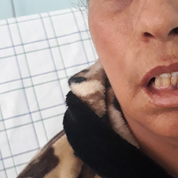
Guillain-Barré syndrome (GBS) is an acute inflammatory polyradiculoneuropathy. Progressive limb weakness, diminished/absent reflexes, sensory disturbance, and variable autonomic dysfunction are its core clinical manifestations. Bifacial weakness with paraesthesias (BFP) is a rare regional variant of GBS and is characterized by simultaneous facial diplegia, distal paraesthesias and minimal or no motor weakness. The association of headache with classic GBS has been rarely reported in the literature, and has not yet been described in the BFP variant. Here we report a misleading case of BFP variant associated with severe headache and mild pleocytosis. The repetition of nerve conduction studies (NCS) was extremely beneficial in this confusing case.
|
Views: 759
HTML: 76
PDF: 335
|
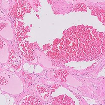
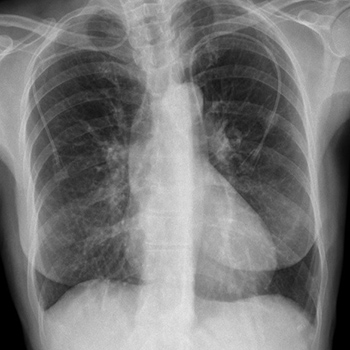
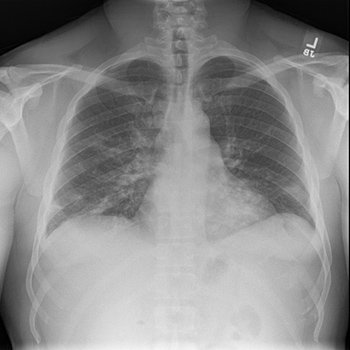
Background: Haemangiomas are uncommon chest wall tumours arising outside the rib cage. Their occurrence in intercostal muscle is extremely rare.
Aim: We describe a case of intercostal muscle cavernous haemangioma as a differential diagnosis for chest wall swelling.
Case description: We describe an 18-year-old male patient with an asymptomatic left-sided chest wall swelling. Contrast-enhanced computed tomography revealed a well-defined homogenously non-enhancing mass lesion arising from the seventh intercostal muscle with differential diagnoses of various chest wall tumours. Clinical presentation and imaging findings were inconclusive, but histopathological examination following excision biopsy revealed a cavernous haemangioma. The present case emphasizes the importance of histopathological diagnosis when clinical and radiological examination is inconclusive. Hence, it is necessary to consider intercostal muscle haemangiomas as a differential diagnosis for chest wall tumours in the absence of a feeding vessel.
Conclusion: Despite its rare occurrence, intercostal muscle haemangioma must be considered as a differential diagnosis in chest wall tumours even in the absence of a feeding vessel. We believe that histopathology can provide a definitive diagnosis when most investigative procedures are inconclusive.
|
Views: 832
HTML: 85
PDF: 450
|
We report a case of delayed diagnosis of cholangiocarcinoma. A 62-year-old man developed acute abdominal pain in multiple sites. As the distribution pattern of the abdominal pain was not correctly interpreted based on the mechanisms of visceral and referred pain, the patient was not investigated with the best diagnostic test at first presentation.
Moreover, miscommunication between physicians in a clinic and separate hospital delayed diagnosis. For prompt diagnosis, physicians should be practice careful reasoning and focus on good communication with physicians outside their hospital.
|
Views: 644
HTML: 72
PDF: 376
|
We describe a 58-year-old Caucasian male weightlifter who presented with acute shortness of breath after finishing his extensive exercise routine. Acute aortic valve regurgitation, due to spontaneous rupture of a bicuspid aortic valve, was diagnosed. Urgent surgical intervention was carried out, during which the bicuspid aortic valve was resected and replaced with an On-X bileaflet mechanical valve. The patient remains asymptomatic and is treated with warfarin, being in excellent physical condition 4 years after aortic valve replacement.

|
Views: 777
HTML: 80
PDF: 390
|
The COVID-19 pandemic has imposed new challenges to scientific community. It’s behaviour and outcomes in people living with HIV is not yet well studied. We report a case of a 34-year-old-male with a newly diagnosed HIV infection stage 4 and asymptomatic SARS-CoV-2 infection. Although immunocompromised patients are classified as a high risk group for developing severe COVID-19, HIV related immunosuppression may have a protective role.
|
Views: 554
HTML: 62
PDF: 359
|
Introduction: Persistence of the left superior vena cava (LSVC) is a rare anatomical variant in the general population with an estimated incidence of 0.3-0.5% in healthy individuals. Its diagnosis can be made incidentally after imaging control of central venous catheter (CVC) or other types of devices placements.
Patient and Methods: We present the case of a patient with an acute disease which required central venous catheterization for the administration of intravenous chemotherapy.
Results: Central venous catheterization proved difficult and after imaging control it revealed an unusual position of the catheter tip. Additional study to verify catheter tip position was performed and computed tomography (CT-scan) revealed the presence of a persistent left superior vena cava. The patient was then submitted to the planned treatment without any record of complications associated with CVC.
Conclusion: Although uncommon the persistence of the LSVC can have an important impact in clinical practice, particularly when more invasive procedures are required. Its recognition is relevant in order to minimize the potential complications inherent to these procedures.
|
Views: 779
HTML: 196
PDF: 413
|
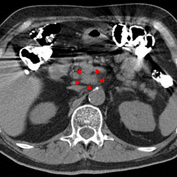
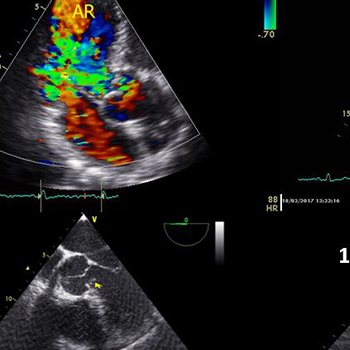
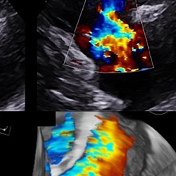
Pancreatic neuroendocrine neoplasms (PanNENs) rarely secrete serotonin, which is the main cause of carcinoid syndrome. One of its unusual manifestations is carcinoid heart disease or Hedinger’s syndrome which is seldom accompanied by cardiac arrhythmias. We report the case of an 88-year-old woman who presented with recently experienced episodes of palpitations and a newly developed atrial flutter with a ventricular rate of 130 beats per minute. Echocardiography revealed thickened and tethered tricuspid and pulmonary valve leaflets causing severe valvular regurgitation and right ventricular dilatation. Episodes of intermittent diarrhoea over the previous 2 years were mentioned, making carcinoid syndrome our working diagnosis. The 5-hydroxyindoleacetic acid (5-HIAA) levels in a 24-hour urine collection specimen were elevated. Conventional imaging studies and a Ga-68 dodecane tetraacetic acid tyrosine-3-octreotate (DOTATATE) positron emission tomography/computer tomography (PET/CT) scan revealed the presence of a metastatic PanNEN arising from the pancreatic tail. The patient was managed with lanreotide and telotristat with remarkable improvement of her symptoms. To our knowledge, this is the first reported case of carcinoid syndrome presenting with atrial flutter as the initial symptom.
|
Views: 937
HTML: 74
PDF: 408
|
Case description: A 64-year-old patient with chronic renal failure and persistent hyperkalaemia not corrected by dialysis, was prescribed sodium polystyrene sulfonate (SPS) at a low dose (30 g/day for 2 days a week during the long interdialytic interval). After 3 months of therapy, the patient developed intense abdominal pain with non-specific colitis identified with a colonoscopy. In addition, the biopsy specimens showed rhomboid SPS crystals in the intestinal mucosa. Fourteen months after discontinuing therapy, the patient again presented with colitis and persistent biopsy finding of SPS crystals. The patient died a few months later due to intestinal infarction.
Discussion and conclusion: SPS is a cation exchange resin used to treat hyperkalaemia resistant to dialysis, but may cause inflammation and ischaemia of the colon. In our patient, a short 3-month course of low-dose SPS therapy (without sorbitol, which is used to counter iatrogenic constipation caused by SPS) induced relapsing colitis, which was followed by massive intestinal infarction a few months later. In light of frequent reports of its enterotoxic effects, SPS should be replaced with the new potassium chelators (patiromer and sodium zirconium cyclosilicate).
|
Views: 558
HTML: 71
PDF: 321
|
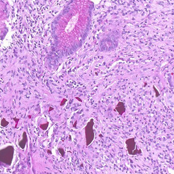
Splenosis is a benign condition which results from the self-implantation of splenic tissue on intra or extraperitoneal surfaces, after splenic trauma or splenectomy. Patients are usually asymptomatic but may present with varied symptoms related to the implantation site. The diagnosis is a challenge because abdominal splenosis can mimic several diseases, including neoplasm. The gold standard examination for its diagnosis is scintigraphy with 99mTc-labelled heat-denatured erythrocyte. When splenosis is found in an asymptomatic patient, surgical removal is not indicated. A 57-year-old male patient presented with sporadic epigastric pain and a suspected mass in the recto-sigmoid transition. Abdominal ultrasound, CT and MRI identified this mass, its characteristics and location, but failed to distinguish its nature. However, given the patient’s past history of splenectomy and because the mass showed a similar sign to that of the splenic parenchyma, a hypothesis of abdominal splenosis was raised, which was confirmed by scintigraphy with 99mTc-labelled heat-denatured erythrocyte.
In this case, the diagnosis was obtained before the patient was subjected to more invasive procedures, which are associated with high morbidity, and, as in most cases, no targeted intervention was necessary.
|
Views: 1355
HTML: 125
PDF: 599
|
Severe acute respiratory syndrome coronavirus 2 (SARS-CoV-2), the virus that causes coronavirus disease 2019 (COVID-19), has caused a global health crisis. COVID-19 can have a multifaceted presentation. A wide range of complications and outcomes can emerge based on the severity and comorbidities of the infected patient. We report a 42-year-old male with past medical history of CML on Dasatinib (in Major Molecular Response) who was diagnosed with COVID-19 and developed pancytopenia. Our case and review of available reports add to the limited literature available regarding COVID-19 in CML.
|
Views: 647
HTML: 90
PDF: 374
|
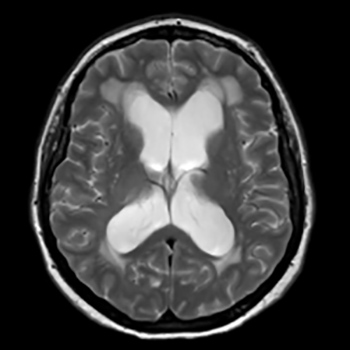
We present the case of a patient with severe obstructive sleep apnoea (OSA) and hypoventilation syndrome who had hydrocephalus and acquired aqueduct stenosis. A link between these conditions in our patient is postulated. We discuss the mechanisms through which this might have occurred and the potential problems which might arise in applying non-invasive ventilation to a patient with hydrocephalus.

|
Views: 1053
HTML: 121
PDF: 572
|
Gitelman syndrome (GS) is a hereditary renal tubulopathy caused by mutations in the SLC12A3 gene which encodes the thiazide-sensitive apical sodium-chloride cotransporter. GS is characterized by hypokalaemia, hypomagnesaemia and metabolic alkalosis. Treatment is based on potassium and magnesium replacement ad eternum. We present the case of a young man with palpitations and persistent hypokalaemia, who was diagnosed with GS. Genetic testing revealed 2 mutations in the gene SLC12A3 of combined heterozygosity, both considered pathological. Interestingly, 1 of these mutations was not yet described in the literature or in the reviewed databases. We also discuss the clinical approach and the specificities of managing this rare hereditary renal tubulopathy.
|
Views: 977
HTML: 725
PDF: 547
|
Ceftriaxone is a widely used antibiotic regarded as safe and effective. Drug-induced agranulocytosis is a life-threatening adverse reaction and few reports related to ceftriaxone were found in a review of the literature. The authors present a case of ceftriaxone-induced agranulocytosis, in which a brain abscess was diagnosed and ceftriaxone was commenced. Neutropenic fever occurred on the 29th day of therapy with a cumulative dose of 116 g ceftriaxone and a neutrophil nadir of 0.1×10⁹/l. Ceftriaxone was withheld, filgrastim was administered for 3 days and neutrophil normalization was achieved. Although rare, ceftriaxone-induced agranulocytosis may occur in patients on a long course of ceftriaxone therapy. Prompt recognition and drug withdrawal are required.
|
Views: 896
HTML: 237
PDF: 553
|
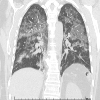
A 57-year-old woman with Crohn's disease (ulcerative proctitis) treated with mesalazine (5-ASA) developed worsening respiratory distress and cough. The lack of response to antibiotics and the results of bronchoalveolar lavage led to the diagnosis of mesalazine-related hypersensitivity pneumonitis, an infrequent entity. Symptoms improved after discontinuation of mesalazine and the administration of corticosteroid therapy. The authors discuss the diagnosis and management of this rare condition.
| 2.1 = | 1.762 Cit. to date |
| 842 Docs. to date |


Publisher
Official Journal of the
European Federation of Internal Medicine
www.efim.org
Publisher: SMC media Srl
Via Giovenale, 7 - 20136 Milan - Italy
P.IVA 07626490960
info@ejcrim.com
www.ejcrim.com - ISSN: 2284-2594 - © EFIM 2014-2024, Published by SMC Media srl, Italy - Privacy policy

