EJCRIM 2023 CiteScore
| 2.1 = | 1.762 Cit. to date |
| 842 Docs. to date |
Last updated on 05 May, 2024
Updated monthly
Updated monthly
Powered by


|
Views: 472
HTML: 763
PDF: 368
|
Well-known side effects of acyclovir are nephrotoxicity and neurotoxicity. We present a 49-year-old woman without pre-existing renal failure, with an acute kidney injury and encephalopathy. Since there was a clear correlation with the intake of acyclovir and the course of illness, findings were attributed to the antiviral agent. Urinalysis showed a proteinuria in nephrotic ranges, which is not described in the currently known causes of acyclovir-induced renal failure. We postulate the hypothesis of a nephritis with podocyte damage induced by acyclovir or, more likely, by an acyclovir metabolite.
|
Views: 453
HTML: 97
PDF: 432
|
Introduction: Guillain-Barré syndrome is an immune-mediated inflammatory polyneuritis characterised by rapidly progressive flaccid paralysis. Guillain-Barré syndrome may present with posterior reversible encephalopathy syndrome or reversible cerebral vasoconstriction syndrome in rare cases.
Case description: A woman in her 60s with a history of follicular lymphoma presented with a one-week history of difficulty walking and thunderclap headaches. The patient was diagnosed with Guillain-Barré syndrome based on neurological examination, cerebrospinal fluid analysis and nerve conduction findings. Further diagnosis of posterior reversible encephalopathy and reversible cerebral vasoconstriction syndromes was based on imaging findings and headache history. The patient was treated with intravenous immunoglobulin and amlodipine, and symptoms improved.
Discussion: We reviewed the literature on Guillain-Barré syndrome associated with posterior reversible encephalopathy and/or reversible cerebral vasoconstriction syndrome. The underlying pathophysiology may involve dysautonomia resulting in unstable blood pressure, and hyponatraemia causing endothelial dysfunction. The SNOOP mnemonic highlights the ‘red flags’. This SNOOP mnemonic suggests the possibility of secondary headaches that require imaging studies. In this case, the patient exhibited three SNOOP symptoms: S (history of malignancy: follicular lymphoma), O (sudden-onset headache) and O (over 50 years old).
Conclusion: This case highlights the importance of considering coexisting central neurological disorders in patients with Guillain-Barré syndrome.
|
Views: 296
HTML: 33
PDF: 259
|
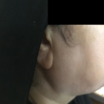
Raoultella ornithinolytica is an encapsulated, Gram-negative, nonmotile, rod belonging to the Enterobacteriaceae family. Infections involving the gastrointestinal tract and the hepatopancreatobiliary system are most frequently reported, especially in immunocompromised patients. The authors present an unusual case of acute complicated sinusitis with orbital and intracranial involvement caused by R. ornithinolytica. The infection was rapidly progressive, even though the patient was a healthy, young person without any co-morbidities. The patient’s condition improved after antibiotic treatment and multiple ophthalmic and sinus surgeries.
|
Views: 258
HTML: 31
PDF: 277
|
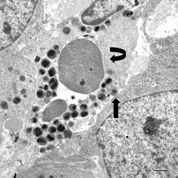
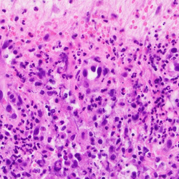
A case of poorly cohesive NOS gastric carcinoma, characterised by high-grade tumour-associated tissue eosinophilia (TATE), is studied by transmission electron microscopy. Eosinophil clustering around single tumour cells constituted a recurrent ultrastructural hallmark. Some eosinophils were in intimate contact with tumour cells and exhibited extracellular trap cell death (ETosis): a non-apoptotic cell death process, recently described in non-neoplastic, eosinophil-associated diseases. Discharge of chromatin material and specific granules, due to eosinophil ETosis, was polarised towards single tumour cells that showed various degrees of cytopathogenic changes. Our data suggest that eosinophil ETosis may exert an antitumoural activity in gastric cancer.
|
Views: 246
HTML: 47
PDF: 257
|
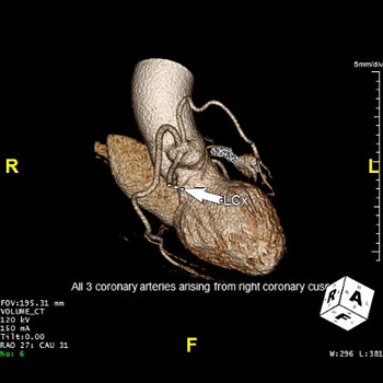
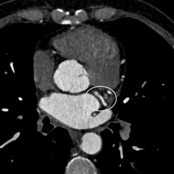
Spontaneous coronary artery dissection (SCAD) is an epicardial coronary artery dissection not associated with atherosclerosis or trauma, and is not iatrogenic. The pathogenesis of SCAD is not fully understood, and the association of coronary artery anomalies and SCAD is not known. We present a case of a 64-year-old woman presenting with non-ST-elevation myocardial infarction (NSTEMI) due to have multivessel SCAD in the setting of anomalous origin of the coronary arteries with all three coronary arteries – right coronary artery (RCA), left anterior descending artery (LAD), and left circumflex artery (LCx) originating from the right sinus of Valsalva.
|
Views: 429
HTML: 30
PDF: 351
|
Hypertrophic pachymeningitis (HP) is an uncommon condition characterised by focal or diffuse thickening of the dura mater. An increasing number of cases have been reported of its association with underlying connective tissue diseases. It is a rare complication in systemic lupus erythematosus (SLE) and might be the initial and sole clinical manifestation. We report a case of a 21-year-old man presenting with febrile meningeal syndrome and sphincter dysfunction. Physical examination showed malar rash and joint pain. Biological assessment revealed a regenerative normocytic normochromic anaemia, a leucopenia and a lymphopenia. The 24-hour urine protein was positive at 0.6 g. Immunological evaluation revealed positive antinuclear, anti-Sm and anti-dsDNA antibodies. Brain and spinal magnetic resonance imaging showed hypertrophic pachymeningitis. Cerebrospinal fluid biochemistry was within normal limits. Renal biopsy revealed a mesangial proliferative lupus nephritis. The diagnosis of SLE with neurologic and renal involvement was established, and the patient was treated with intravenous methylprednisolone pulse, followed by oral prednisone in association with azathioprine and hydroxychloroquine. Considering the persistence of symptoms and MRI lesions after 6 months, a treatment with rituximab was initiated with good evolution.
|
Views: 467
HTML: 71
PDF: 342
|
Infectious mononucleosis (IM), the most common presentation of acute Epstein Barr virus (EBV) infection, typically presents with fever, pharyngitis and lymphadenopathy. We describe an unusual case of IM presenting as acute sinusitis. A 25 year-old male presented to the emergency department with worsening right frontal sinus pain along with fever, chills, and greenish nasal discharge for 3 weeks. Laboratory workup showed leukocytosis with high lymphocyte counts as well as transaminitis. Facial computerized tomography (CT) showed extensive right frontal, ethmoidal and maxillary sinusitis and antrochoanal polyp. The patient underwent endoscopy with drainage of purulent material and polyp removal. Unfortunately, cultures of the sample were not sent and bacterial infection could not be ruled out. Broad spectrum antibiotics were continued. Pathology of redundant tissue revealed large atypical lymphocytes with positive EBV-encoded RNA and lack of evidence of extranodal natural killer/T-cell (NK/T-cell) type lymphoma (ENKTCL). Tests for serum EBV IgM antibodies and EBV early Antigen antibodies were positive, indicating acute EBV infection. Lymphocytosis resolved along with significant clinical improvement at the 10-day follow up visit. Even though patient did receive antibiotics, multiple factors including isolated lymphocytosis, pathology positive for EBV with no neutrophilia were more suggestive of sinusitis caused by viral infection, EBV in this case. Lymphocytosis with fever and sore throat should prompt physicians to consider IM. There are no known reports in the literature of EBV as a causal organism for acute viral sinusitis. There are some studies relating EBV with ENKTCL. It is unknown whether this particular patient with a history of EBV sinusitis will be at high risk for nasal type lymphoma in the future. Further studies should be conducted to understand the pathogenesis and relationship between EBV and ENKTCL.
|
Views: 541
HTML: 99
PDF: 571
|
Background: Linezolid is known to cause side effects, including nausea, diarrhea, and headaches of short duration. As extended use of linezolid is becoming more common, additional rare side effects should be considered.
Case Presentation: A 68-year-old man hospitalized for osteomyelitis developed severe abdominal pain and altered mental status following five weeks of linezolid therapy. Laboratory studies showed very high lipase levels, lactic acidosis not responding to resuscitation, and relative hypoglycemia. All common causes of pancreatitis were ruled out, and a trial of linezolid withdrawal was done resulting in drastic improvement in the patient's clinical status.
Conclusions: For patients on extended course of linezolid who develop abdominal pain, drug-induced pancreatitis should be considered as a side effect, and a trial of withdrawal of linezolid should be undertaken.
|
Views: 466
HTML: 72
PDF: 405
|
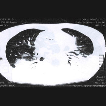
Introduction: Organising pneumonia belongs to diffuse interstitial lung diseases; we distinguish the cryptogenic organising pneumonia, which is idiopathic, from the secondary organising pneumonia caused by drugs or a defined cause. Denosumab is a human monoclonal antibody, rarely inducing adverse pulmonary effects.
Case description: A 57-year-old female patient was admitted to our chest clinic for acute respiratory distress. She was treated with denosumab for severe osteoporosis. The patient described a dry cough and dyspnoea over the previous four months, increased after the last injection of denosumab. A high-resolution computed tomography scan showed bilateral basal parenchymal condensations. The aetiological investigation did not reveal any infectious or immunological origin. The favourable computed tomography imaging and clinical evolution after corticosteroid therapy led to the diagnosis of drug-induced organising pneumonia.
Conclusion: Denosumab could induce organising pneumonia. Therefore, clinicians should be aware of this pulmonary toxicity.
|
Views: 356
HTML: 43
PDF: 327
|
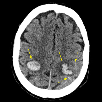
Introduction: Intracranial haemorrhage may complicate infective endocarditis, caused by ruptured mycotic aneurysms or haemorrhagic transformation of brain septic emboli. The risk of intracranial bleeding may increase with the use of non-steroidal anti-inflammatory agent (NSAIDs).
Case description: We report on a 53-year-old male patient with a past history of intravenous drug abuse, who was treated with diclofenac (75 mg IM) for a few hours of preceding fever and arthralgia. Seven hours later he was hospitalised with impaired consciousness and hemiparesis. Evaluation revealed multiple intracranial haemorrhages, at least one originating from a mycotic aneurysm. Repeated blood cultures grew methicillin-resistant Staphylococcus aureus (MRSA), and echocardiography revealed a vegetation on the mitral valve, establishing the diagnosis of bacterial endocarditis.
Conclusion: The abrupt simultaneous multifocal intracranial bleeds shortly following the administration of NSAIDs for a few hours of febrile disease, one clearly originating from a mycotic aneurism, are exceptional. This raises a possibility of a role for diclofenac the intracranial bleeding diathesis in this unique clinical presentation. Intracranial haemorrhage in the set-up of undiagnosed infective endocarditis (IE) might be added to the long list of potential adverse outcomes of NSAID administration, and the possibility of IE should be considered before their administration for febrile disease of undetermined cause.
|
Views: 270
HTML: 124
PDF: 249
|
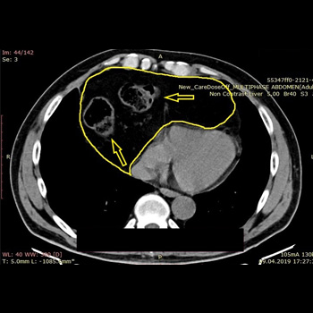
Background: Congenital diaphragmatic hernias are rare congenital defects resulting in abdominal organ protrusion into the thoracic cavity; they mainly present with pulmonary or gastrointestinal symptoms. Although congenital and discovered in utero or in early childhood, they can be asymptomatic for a long time and even remain asymptomatic despite the growing hernia sac dimensions and the hernia sac contents.
Case description: We present a case of a 58-year-old patient with incidentally diagnosed Morgagni hernia during the COVID-19 pandemic following a computerised tomography (CT) scan of the chest. He presented without any symptoms related to the existence of the hernia. Another CT scan was performed 20 months after the initial diagnosis to evaluate the progression of the hernia. The patient refused the offered surgery due to the absence of symptoms.
Discussion: A Morgagni hernia is usually discovered during pregnancy or in early childhood, but sometimes can be asymptomatic for years. Main symptoms originate from the respiratory and gastrointestinal system.
Conclusion: Due to the refusal of surgery, we were able to follow the CT scan enlargement progression of patients’ hernia over a 20-month period.
|
Views: 217
HTML: 115
PDF: 210
|
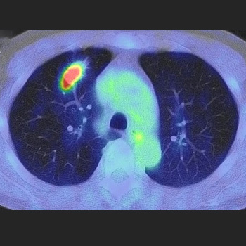
A 69-year-old man was diagnosed with lung adenocarcinoma with metastasis because two masses in the right intercostal space and right back muscle showed high accumulation on positron emission tomography (PET). The 6-month treatment with osimertinib significantly reduced his lung lesion, but no changes were observed in the metastatic lesions. Needle biopsy revealed that the lesion in the right back muscle was a schwannoma. Surgical resection revealed that the right intercostal lesion was also a schwannoma; subsequently, a right upper lobectomy was performed. The patient was finally diagnosed with lung adenocarcinoma without metastasis. High accumulations of lesions observed on PET may indicate schwannomas.
|
Views: 611
HTML: 845
PDF: 681
|
Introduction: Primary Sjögren syndrome (pSS) is an immune systemic disease, that may affect the central nervous system. A severe headache unresponsive to treatment is the headache which is persistently nonresponsive to narcotic analgesics.
Case presentation: A 48-year-old woman with a 10-year history of pSS was seen in January 2021, complaining of a headache one week previously. The headache was characterised by a dull persistent pressing intensity and was not responding to paracetamol, NSAIDs or codeine. She had no previous history, nor family history. Physical examination revealed bilateral parotid glands enlargement. Laboratory tests showed anaemia, and elevated levels of erythrocyte sedimentation rate (ESR) and C-reactive protein (CRP), with positive anti-La and anti-Ro antibodies. She was given topical treatment and different doses of Predlone, in addition to methotrexate10 mg/week. She had received three pulses of methylprednisolone and was started on azathioprine with a mild response to the headaches, so she received two initial IV doses of rituximab 375 mg/m2, then every 2 weeks, with a clinical and laboratory response. Two years later, she had no headache.
Discussion: Headache that may presented in pSS are tension headaches, migraines and cluster headaches. The therapy is disease-modifying antirheumatic drugs, hydroxychloroquine, glucocorticoids and biotherapeutics. Rituximab is used in the treatment of some patients with pSS, especially where it can affect systemic symptoms.
Conclusion: Rituximab treatment may be an option for severe headache in patients with pSS. The mechanism is unknown but may be due to depletion of brain auto-reactive B cells. Further research is needed.
|
Views: 334
HTML: 44
PDF: 307
|
Coronary artery fistulas (CAFs) are rare defects in the coronary circulation system that are usually diagnosed incidentally with cardiac imaging. Although the prognosis of coronary artery fistulas is highly variable, the complications to which they predispose patients are ultimately the determining factor. The authors describe a case of a 56-year-old male, a smoker, hospitalised for worsening dyspnoea on progressively smaller efforts, in the context of acute heart failure. During hospitalisation and imaging exams, a coronary-bronchial fistula was identified.
|
Views: 312
PDF: 405
HTML: 567
|
Introduction: Behçet's disease is a systemic vasculitis characterized by a large clinical polymorphism with a particular frequency of cutaneous signs. Sweet's syndrome is a neutrophilic dermatosis marked by the sudden appearance of painful skin lesions in the form of erythematous papules, nodules or plaques. This syndrome is associated with high fever, neutrophilia and histologically a diffuse infiltrate of neutrophils in the dermis.
Observation: We report the case of a 43-year-old patient followed for Behçet's disease, who developed cutaneous plaques of neutrophilic dermatosis of both upper limbs. The clinical and biological picture was in favor of Sweet's syndrome.
Conclusion: The coexistence of Sweet's syndrome and Behçet's disease is already reported in the literature. The association is however very rare given the differences in the clinical and pathogenic features between the two conditions.
| 2.1 = | 1.762 Cit. to date |
| 842 Docs. to date |


Publisher
Official Journal of the
European Federation of Internal Medicine
www.efim.org
Publisher: SMC media Srl
Via Giovenale, 7 - 20136 Milan - Italy
P.IVA 07626490960
info@ejcrim.com
www.ejcrim.com - ISSN: 2284-2594 - © EFIM 2014-2024, Published by SMC Media srl, Italy - Privacy policy

