EJCRIM 2023 CiteScore
| 2.1 = | 1.762 Cit. to date |
| 842 Docs. to date |
Last updated on 05 May, 2024
Updated monthly
Updated monthly
Powered by


|
Views: 965
HTML: 149
PDF: 459
|
Objectives: This is the first case report of iatrogenic Takotsubo syndrome (TS) due to a combination of lisdexamfetamine and phentermine.
Background: TS is characterized by transient acute ballooning of the left ventricular wall. Typically, it occurs in extremely stressed post-menopausal women, however a few iatrogenic causes have been described recently.
Results: A 55-year old woman prescribed lisdexamfetamine and phentermine, presented with acute substernal chest pain. Acute coronary syndrome was excluded. The echocardiogram was diagnostic of TS, and she recovered spontaneously, with supportive care.
Conclusion: Caution with the use of sympathomimetic medications in post-menopausal women appears warranted.
|
Views: 964
HTML: 122
PDF: 452
|
Introduction: Twenty-five per cent of tuberculosis patients have pleural tuberculosis, which is the third most common form of presentation. Most cases present as an exudative pleural effusion with just few cases reported as chylothorax in the literature. All pleural effusions from confirmed cases, including tuberculous chylothorax, had exudate features.
Aim: To describe a patient with Mycobacterium tuberculosis affecting the lungs and pleura, which laboratory testing demonstrated had features of transudate chylothorax.
Patient and methods: A 70-year-old man presented with constitutional symptoms, progressive exertional dyspnoea and right pleural effusion with fibrocavitary changes on chest imaging. Thoracentesis and pleural fluid analysis revealed chylous fluid with transudate features, high triglycerides, low cholesterol content and mononuclear cell predominance. Acid-fast sputum stains and pleural fluid were negative for Mycobacterium tuberculosis as was an adenosine deaminase test for pleural effusion. Tomography-directed lung biopsy sampling of a lung nodule revealed a chronic granulomatous inflammatory process associated with the presence of acid-fast bacilli.
Discussion: Tuberculosis-associated chylothorax is an uncommon presentation of the disease. A recent review found only 37 cases of confirmed tuberculous chylothorax had been reported in the literature. All cases had exudate characteristics. The diagnosis of pleural tuberculosis was made through culture or testing of sputum, pleural fluid or biopsy samples in 72.2% of cases, with the rest identified by histopathology.
|
Views: 772
HTML: 70
PDF: 333
|
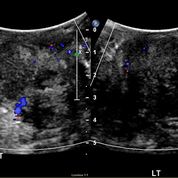
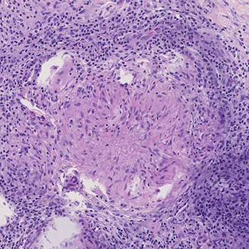
In the present report, we describe our experience with a 44-year-old male with abnormal retroperitoneal primitive neuroectodermal tumours (PNETs) in our hospital, who was operated on with a spindle cell neoplasm diagnosis.
|
Views: 1246
HTML: 91
PDF: 556
|
Despite a recent decline, tuberculosis (TB) infection is still a frequent diagnosis in Portugal. Adenosine deaminase (ADA) measurement has become an important tool in the timely diagnosis of this infection. However, ADA elevation in bodily fluids is not pathognomonic of TB infection.
We present the case of a 70-year-old woman, undergoing treatment for pleural TB, diagnosed based on elevated ADA levels in a pleural effusion. Due to worsening symptoms she was readmitted, and the previous diagnosis was reconsidered. Thoracocentesis was repeated and cytometry analysis of the fluid was performed, showing the presence of diffuse large B cell lymphoma (DLBCL).
DLBCL is the most frequently occurring non-Hodgkin lymphoma (NHL). Pleural involvement is rare in the initial stages. ADA elevation >250 U/l should raise suspicion of malignancy, especially in association with markedly elevated LDH levels. The purpose of this case report is to highlight that in the absence of microbiologic or histologic confirmation, a presumptive TB diagnosis should not be lightly made, and alternative diagnoses should be systematically ruled out.

|
Views: 828
HTML: 146
PDF: 645
|
While functional decline is a common syndrome in geriatric medicine, the diagnosis of the underlying disease can be complex. We present a case of very late-onset systemic lupus erythematosus with fever, arthritis, lymphadenopathy, sicca syndrome, pleurisy, renal impairment and reversible functional and cognitive impairments. Prompt improvement was observed on prednisolone and hydroxychloroquine.
|
Views: 795
HTML: 80
PDF: 355
|
Testicular adrenal rest tumour (TART) is a known entity in patients with congenital adrenal hyperplasia. An adult patient presenting with testicular enlargement raises a concern for malignancy and this creates a diagnostic dilemma between non-malignant conditions such as TART versus testicular malignancy. We describe a case where the patient underwent orchiectomy due to clinical concern for malignancy but, retrospectively, this outcome could have been prevented by medical treatment. This case emphasises the need to learn from errors. There is a need to increase awareness of the condition among medical professionals to reduce the chances of unnecessary surgical intervention.
|
Views: 833
HTML: 233
PDF: 389
|
Case presentation: A 50-year-old female presented with an onset of multiple subcutaneous nodules on her 4 limbs. These nodules appeared concomitantly with the initiation of radioactive iodine therapy for papillary thyroid cancer. These nodules were not obvious on inspection of the skin, but easily felt on palpation.
The biopsy of the subcutaneous nodules revealed hypodermic non-caseating granulomas consistent with sarcoidosis. The patient underwent an 18F-fluorodeoxyglucose positron emission tomography (PET) scan study that revealed, besides the subcutaneous nodules, multiple hypermetabolic mediastinal lymphadenopathies and cervical adenopathies. Biopsy of the mediastinal lymphadenopathy showed neither granulomas nor neoplastic cells. Cervical biopsy revealed neoplastic cells of thyroid origin. Laboratory tests were normal. Bronchoalveolar lavage showed a normal CD4/CD8 T-cell ratio.
A diagnosis of cutaneous sarcoidosis was established, as well as a recurrence of the cancerous disease. The subcutaneous nodules regressed spontaneously in the absence of any treatment.
Discussion and conclusion: Sarcoidosis is a multisystemic disease of unknown origin. This case illustrates an uncommon occurrence of sarcoidosis, triggered by radioactive iodine therapy. Radioiodine may lead to immunological changes, especially affecting the Th1/Th2 ratio, which may promote the emergence of sarcoidosis in genetically predisposed patients. There is still much to discover to fully understand the pathogenesis of sarcoidosis.

|
Views: 805
HTML: 157
PDF: 386
|
A 26-year-old woman presented with a 3-month history of worsening episodic abdominal pain, which was associated with frequent passage of watery stools, nausea and dyspepsia. Her peripheral eosinophil count was markedly elevated. This responded well to a reducing regimen of corticosteroids. Her symptoms completely resolved with a corresponding fall in eosinophil count. The patient was diagnosed with eosinophilic gastroenteritis. We have not considered steroid-sparing agents at this point, but should she have future exacerbations then this will be considered.
|
Views: 789
HTML: 116
PDF: 421
|
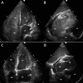
Background: Right heart thrombus (RHT) is a medical condition associated with acute pulmonary embolism and congestive cardiac failure. Rapid recognition is essential for instituting early treatment and preventing adverse outcomes.
Case summary: A 55-year-old male presented with symptoms of congestive cardiac failure complicated by cardiac arrest. Initial transthoracic echocardiography (TTE) demonstrated moderate impairment of both ventricles and a moderately dilated right ventricle (RV). After initial improvement with heart failure treatment, the patient subsequently had a second cardiac arrest. Bedside TTE revealed complete RV obstruction by thrombus, and intravenous thrombolysis was immediately instituted, with complete dissolution of the thrombus and haemodynamic recovery 15 minutes after treatment. Unfortunately, the patient suffered significant hypoxic brain injury and did not survive.
Discussion: RHT can manifest acutely in a dramatic fashion with cardiac arrest. Bedside TTE is key to making a rapid diagnosis in this setting to allow early administration of thrombolytic therapy.

|
Views: 945
HTML: 404
PDF: 512
|
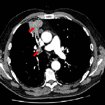
Aortoenteric fistula (AEF) is a rare condition with a high mortality rate. AEFs are classified according to their primary and secondary causes, the former being less frequent. Primary AEFs occur in a native aorta and their causes include aneurysms, foreign bodies, tumours, radiotherapy and infection. The classic triad of aortoesophageal fistulas, a subtype of AEFs, are mid-thoracic pain and sentinel haemorrhage, followed by massive bleeding after a symptom-free interval.
We present the case of a 41-year-old male patient who presented in the emergency room after successive episodes of abundant haematemesis. He was hypovolemic, hypothermic and acidotic at presentation. His medical history included an emergency room visit the week before with chest pain but no relevant anomalies on work-up, active intravenous drug use and chronic hepatitis.
Esophagogastroduodenoscopy (EGD) showed a bulging ulcerated lesion suspicious for aortoesophageal fistula, confirmed by computed tomography (CT) angiography, which revealed a saccular aortic aneurysm with a bleeding aortoesophageal fistula. The patient underwent urgent thoracic endovascular aortic repair.
The sentinel chest pain, leucocytosis and CT findings hinted at the presence of a mycotic aneurysm, despite the negative blood cultures. It was most likely caused by a septic embolus due to the patient’s risk factors. While a high level of suspicion for aortoesophageal fistula is needed to prompt a fast diagnosis, EGD and CT findings were crucial to establish it and allow a life-saving intervention. We conclude that chest pain cannot be disregarded in a patient aged 41 years with multiple comorbidities, despite normal work-up, to prevent a fatal outcome.
|
Views: 1769
HTML: 87
PDF: 449
|
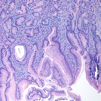
A 77-year-old man with arterial hypertension and dyslipidaemia, treated with olmesartan/hydrochlorothiazide and simvastatin, was admitted with a 3-week history of anorexia, nausea, vomiting, profuse diarrhoea and weight loss. He was dehydrated and blood tests showed acute kidney injury. The aetiological study was inconclusive. The patient had a favourable clinical evolution during hospitalization and was discharged. However, after about 10 days at home, he was re-admitted to hospital with the same clinical presentation. It was noticed that olmesartan had not been prescribed during the previous admission but had been restarted on an outpatient basis. Biopsy examination showed duodenal mucosa with villous atrophy and polymorphic inflammatory infiltrate. Antibody testing for coeliac disease was negative. Based on these facts, it was hypothesized that the patient had olmesartan-induced enteropathy, which was subsequently confirmed.
|
Views: 864
HTML: 125
PDF: 501
|
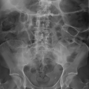
Ogilvie’s syndrome is a non-mechanical, acute pseudo-obstruction of the colon, causing massive colonic dilation. Medical or surgical conditions can predispose patients to Ogilvie’s syndrome; however, the pathogenesis and clinical findings are still not well understood. Here, we present a case of a 48-year-old male patient who presented to the Emergency Department with intermittent self-resolved left-sided lower chest pain on a background of ischaemic heart disease and positive risk factors for acute coronary syndrome. Troponin testing was negative and an electrocardiogram showed no acute changes. Chest radiography showed a dilated bowel under the left hemidiaphragm and a computed tomography (CT) scan of the abdomen-pelvis confirmed the diagnosis of Ogilvie’s syndrome. The patient was treated conservatively with a short period of nil by mouth and intravenous fluids.

|
Views: 690
HTML: 77
PDF: 505
|
Acquired haemophilia (AHA) is a rare autoimmune disorder caused by circulating autoantibodies that inhibit the activity of factor VIII (FVIII). Acquired inhibitors against FVIII are rarely seen, with a reported incidence of approximately 1 case per million/year. Clinical conditions and contexts associated with AHA include autoimmune diseases, lymphoproliferative malignancies, drug treatment, pregnancy and infections. An association with urticarial vasculitis is even more rare. Here, we report a case of a 59-year-old woman presenting with cutaneous and muscle haematomas secondary to AHA in association with urticarial vasculitis, who was successfully treated with factor eight inhibitor bypassing activity (FEIBA) and prednisolone.
|
Views: 766
HTML: 69
PDF: 385
|
Background: Coccidioidomycosis is an endemic disease in the Americas. No cases have been reported in Africa.
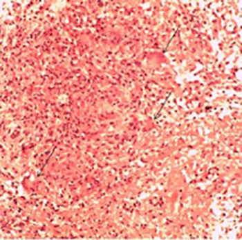
Patient: A 23-year-old HIV seronegative Ugandan man was referred to Mulago National Referral Hospital in Kampala, Uganda with a 10-month history of haemoptysis and difficulty breathing, and a 6-month history of localized swellings on the extremities. He had associated weight loss and drenching sweats, but no fevers. He had taken anti-tuberculosis medicine for 2 months with no improvement. He had never travelled out of Uganda. On physical examination, he had cystic swellings and ulcerated lesions on the extremities. He had tachypnoea, crackles in the chest and mild hepatomegaly. Bronchoscopic examination showed two masses occluding the right main bronchus. Bronchoscopic biopsy showed findings consistent with coccidioidomycosis. The patient improved with antifungal treatment and was discharged.
Conclusion: We report the first case of disseminated coccidioidomycosis with pulmonary and cutaneous manifestations in Africa.
|
Views: 1062
HTML: 1546
PDF: 754
|
The lifetime prevalence of peptic ulcer disease (PUD) is 5–10%. While PUD in immunocompetent patients is most commonly associated with Helicobacter pylori infection or the use of non-steroidal anti-inflammatory drugs (NSAIDs), other common causes of PUD must also be considered in the differential diagnosis. We describe a case of endoscopic and histological resolution of PUD related to Candida infection in a healthy, immunocompetent woman.
|
Scleroderma with Acro-Osteolysis and Papular Mucinosis Resembling Multicentric Reticulohistiocytosis
Views: 678
HTML: 136
PDF: 395
|
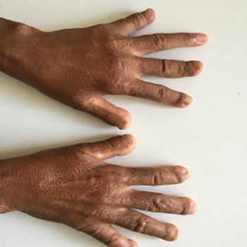
Objectives: We describe a case of systemic sclerosis (SS) with acro-osteolysis associated with cutaneous mucinosis, usually characterized by mucin deposition in the skin. The main differential diagnosis was multicentric reticulohistiocytosis due to the presentation of papulonodular skin lesions.
Materials and methods: A physical examination, imaging studies and laboratory tests were performed.
Results: Distal bone resorption was evident on plain radiographs, and skin biopsy confirmed mucinosis. The SS diagnosis was based on the clinical features, high levels of antinucleolar antibodies and typical nailfold capillaroscopy findings.
Conclusion: To the best of our knowledge, this is the first description of cutaneous mucinosis accompanying SS with acro-osteolysis.
|
Views: 762
HTML: 81
PDF: 344
|
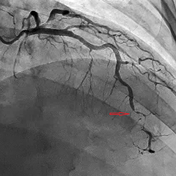
Spontaneous coronary artery dissection (SCAD) is a rare cause of acute coronary syndrome (ACS). Although uncommon, it should be included in the differential diagnosis for middle-aged patients without elevated atherosclerotic vascular disease risk or a family history of cardiovascular disease. SCAD is associated with postpartum women; however, reports noting its association with autoimmune disease and vasculopathy in other populations have recently gained prominence. We report a case of a 41-year-old male who was found to have SCAD after presenting with ST segment elevation myocardial infarction in the context of episodic vision loss, and who later underwent work-up for C-ANCA vasculitis and was successfully treated with corticosteroids.
|
Views: 612
HTML: 456
PDF: 298
|
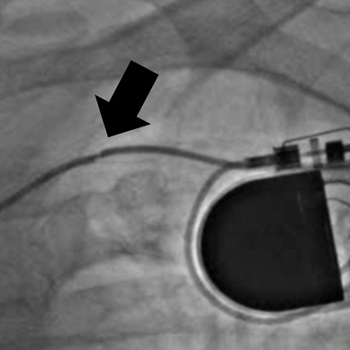
Subclavian vein access is still one of the most favoured access options for cardiac implantable electronic device (CIED) implantation. For the physician, the technique is reasonably familiar and easy to carry out. However, this has several potential complications. In this case, we present a late complication of subclavian access. The patient presented with intermittent loss of pacemaker output, which caused him to experience several syncopal events. In the acute setting, we changed the lead polarity and achieved a good outcome. Further management of this situation consisted of removal and replacement of the damaged lead.
|
Views: 1072
HTML: 116
PDF: 380
|
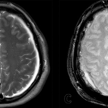
Obstructive sleep apnoea (OSA) is a common condition usually treated with continuous positive airway pressure (CPAP). No reports have linked it to an acute subdural haematoma. A 54-year-old white man who had hypertension well controlled with an angiotensin II receptor blocker, presented with a 2-week history of occipital headache with no other focal neurological symptoms. The headache began 12 days after he had started using CPAP for OSA. A brain MRI performed 2 weeks later showed bilateral subdural haematomas which were chronic on the left and sub-acute/acute on the right. Since the patient was clinically stable with no focal neurological deficits, he received prednisone for 3 weeks and was followed up with consecutive CT scans demonstrating gradual regression of the haematomas. This is the first report showing that subdural haematomas could be linked to CPAP use.
|
Views: 905
HTML: 523
PDF: 424
|
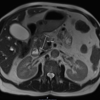
Objective: We present a patient with pancreatic cancer who developed weakness, acute renal failure and significantly raised creatine kinase levels post-ERCP and who was assessed as having contrast-induced rhabdomyolysis.
Results: The patient underwent haemofiltration and ultimately succumbed to his condition.
Conclusion: Rhabdomyolysis is a potentially life-threatening condition which occurs because of damage to skeletal muscle, with release of myoglobin and electrolytes into the circulation. The mortality rate is 59% in severe cases, despite appropriate treatment.
|
Views: 611
HTML: 61
PDF: 313
|
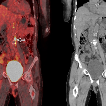
Objectives: We describe the novel case of a patient presenting with pulmonary mucosa-associated lymphoid tissue lymphoma (pMALToma) synchronous with metastatic prostate adenocarcinoma.
Materials and methods: We report the clinical, laboratory, radiological and histological findings of the above patient.
Results: While the patient’s metastatic prostate adenocarcinoma responded well to chemo-radio-hormonal therapy, a persistent area of lung consolidation was noted and further investigated, leading to the diagnosis of concurrent pMALToma.
Conclusion: It is important to pursue further investigation when there appears to be persistent change or altered disease response in malignancy if there is evidence for disease response elsewhere, as there may be two synchronous primary cancers.
|
Views: 911
HTML: 155
PDF: 685
|
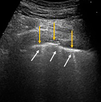
Clinical experience and scientific articles have shown that patients infected with severe acute respiratory syndrome coronavirus 2 (SARS-CoV-2) can be paucisymptomatic or asymptomatic at the time of diagnosis. In this paper, we will discuss two paucisymptomatic patients with blood tests suggestive for SARS-CoV-2 infection but with repeated negative nasopharyngeal swabs and without typical features of COVID-19 pneumonia on chest high-resolution computed tomography. In these cases, lung ultrasound helped to raise clinical suspicion of COVID-19 pneumonia and facilitate diagnosis.

|
Views: 1430
HTML: 136
PDF: 580
|
The ongoing outbreak of coronavirus disease 2019 (COVID-19) that began in Wuhan, China, became an international emergency when thousands of people were infected around the world. COVID-19 emerged in Pakistan in April 2020, precipitating a nationwide lockdown. While some countries are now recovering from the pandemic, its peak is not estimated to occur in Pakistan until August 2020. We present a case of rheumatic heart disease with fever, myalgia and an unusual radiological finding of the virus.

|
Views: 935
HTML: 297
PDF: 476
|
It is increasingly recognised that patients with severe COVID-19 infection have a significant risk of thromboembolic events. We describe a patient who rapidly deteriorated due to severe infection with COVID-19, and developed priapism in the last days of his life. We believe development of priapism may be associated with a prothrombotic state secondary to COVID-19 infection. This case report supports the widely reported increased incidence of thrombosis in patients with severe COVID-19 infection.
|
Views: 1289
HTML: 116
PDF: 606
|
The SARS-CoV-2 virus is a newly emergent pathogen first identified in Wuhan, China, and responsible for the COVID-19 global pandemic. In this case report we describe a manifestation of non-bacterial thrombotic endocarditis with continuous peripheral embolization in a COVID-19-positive patient. The patient responded well to high-dose LMWH treatment with cessation of the embolic process.
|
Views: 1602
HTML: 149
PDF: 688
|
Pityriasis rubra pilaris (PRP) is a rare chronic inflammatory papulosquamous dermatosis affecting both adults and children. Six subtypes of PRP have been described. Recently, the management of PRP with biologic immunosuppressive agents regularly used in psoriasis has been supported by several case reports and series. Ustekinumab is an anti-IL12/23 IgG1 kappa human monoclonal antibody. It has been approved for the treatment of Crohn’s disease, plaque psoriasis, psoriatic arthritis and ulcerative colitis. It has also been reported to be effective as an off-label treatment for PRP. Current data are equivocal regarding infectious disease risk with ustekinumab administration. We describe a case of meningococcal and HSV-2 infection of the central nervous system in a patient being treated with ustekinumab for PRP.
| 2.1 = | 1.762 Cit. to date |
| 842 Docs. to date |


Publisher
Official Journal of the
European Federation of Internal Medicine
www.efim.org
Publisher: SMC media Srl
Via Giovenale, 7 - 20136 Milan - Italy
P.IVA 07626490960
info@ejcrim.com
www.ejcrim.com - ISSN: 2284-2594 - © EFIM 2014-2024, Published by SMC Media srl, Italy - Privacy policy

