EJCRIM 2023 CiteScore
| 2.1 = | 1.730 Cit. to date |
| 842 Docs. to date |
Last updated on 05 April, 2024
Updated monthly
Updated monthly
Powered by


|
Views: 976
HTML: 99
PDF: 426
|
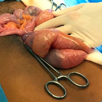
Meckel’s diverticulum, a congenital malformation of the gastrointestinal tract, is asymptomatic in the majority of patients but can be associated with some complications. Gastrointestinal bleeding is one such complication and is more common in children than in adults. Despite the variety of examinations available, diagnosis can be difficult, especially in older patients, because the sensitivity of examinations decreases with patient age. Here we present the case of a young man with gastrointestinal bleeding in whom a diagnosis of Meckel’s diverticulum was made intra-operatively.
|
Views: 1237
HTML: 297
PDF: 529
|
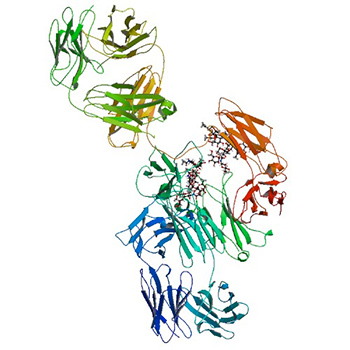
Amyloidosis is a group of disorders characterised by the accumulation of extracellular deposits of insoluble protein aggregates. Clinical management depends on the accurate identification of the amyloid precursor and underlying cause. We describe a rare case of apolipoprotein A-IV cardiac amyloidosis, the diagnosis of which required mass spectrometry-based proteomic analysis.
|
Views: 1205
HTML: 85
PDF: 434
|
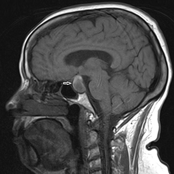
Pituitary apoplexy is a rare medico-surgical emergency that stems from an acute expansion of a pituitary adenoma from infarction or haemorrhage and where the treatment strategy is still controversial. Clinical presentation is highly variable and a high index of suspicion is needed to make the diagnosis. Furthermore, in less than half of cases, a precipitating event is identified. We report a case of a 74-year-old female who, after introduction of anticoagulation for pulmonary thromboembolism, presented with pituitary apoplexy heralded by acute adrenal insufficiency, headaches, visual symptoms and hypogonadotropic hypogonadism. Timely initiation of corticosteroids was crucial, and after stabilisation, a conservative treatment strategy was favoured with good long-term prognosis. Long-term follow-up of pituitary function also revealed new growth hormone deficiency.
|
Views: 1156
HTML: 79
PDF: 572
|
Iatrogenic antineutrophil cytoplasm antibody (ANCA)-associated vasculitis (AAV) is not exceptional. Many cases of small vessel vasculitis induced by anti-thyroid drugs (ATD), mainly propylthiouracil (PTU), have been reported. We present a case of AAV related to another ATD: benzylthiouracil (BTU) and review the literature. An 84-year-old man with a 4-year history of multinodular goitre with hyperthyroidism was treated with BTU. He presented an acute syndrome with weakness, fever, epigastric pain and abdominal distension. Lactate and lipase tests were normal. An abdominal scan showed a thrombosis of the splenic artery with splenic infarction. We excluded a hypothesis of associated embolic aetiology: atrial fibrillation, atrial myxoma, intraventricular thrombus or artery aneurysm. Exploration of a possible prothrombotic state (complete blood count, haemostasis tests, activated protein C resistance, factor V Leiden, protein C, S, antithrombin III) gave normal results. Tests for antinuclear antibodies (ANA) and antiphospholipid antibodies (APL) were negative. However, testing for p-ANCA, with antimyeloperoxidase (MPO) specificity, was positive: 120.6 CU (N<20.0). We did not find other systemic manifestations, except a non-specific kidney failure. BTU was discontinued without steroids or immune-modulating drugs. Subsequently, symptoms disappeared progressively and titres of ANCA fell until normalization, 4 months later. Many patients treated with BTU present a high prevalence of ANCA, mainly, but not exclusively, directed against MPO. Vasculitis, however, remains an uncommon complication. The mechanism of this anomaly remains to be elucidated. Some studies suggest the possibility of an autoimmune reaction initiated by drug bioactivation mediated by neutrophil-derived MPO. The present observation is particular because the involved drug was BTU and clinical expression was unusual.
|
Views: 914
HTML: 90
PDF: 381
|
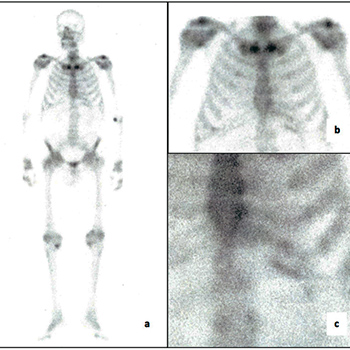
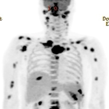
Fluorine-18-fluorodeoxyglucose positron emission tomography/ computed tomography (18FDG-PET/CT) has been used to diagnose vasculitis, tuberculosis and malignancy. Because PET/CT scan show hotspots during an activation of clinically suspected lesions, it is widely used for diagnosis. However, there are rare cases of PET/CT images for vasculitis combined with tuberculosis. Here we report a case of an eosinophilic granulomatosis with polyangiitis in a patient with disseminated non-tuberculosis mycobacterial infection in multiple sites mimicking metastatic malignacy and describe the associated PET/CT scan findings before and after treatment.
|
Views: 922
HTML: 478
PDF: 374
|
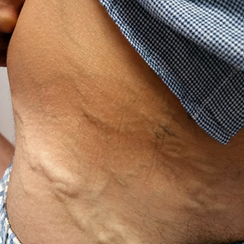
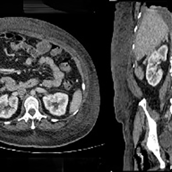
Inferior vena cava (IVC) agenesis is a rare congenital abnormality affecting the infrarenal segment, the suprarenal or the whole of the IVC. It has an estimated prevalence of up to 1% in the general population that can rise to 8.7% when abnormalities of the left renal vein are considered. Most IVC malformations are asymptomatic but may be associated with nonspecific symptoms or present as deep vein thrombosis (DVT). Up to 5% of young individuals under 30 years of age with unprovoked DVT are found to have this condition. Regarding the treatment of IVC agenesis-associated DVT, there are no standard guidelines. Treatment is directed towards preventing thrombosis or its recurrence. Low molecular weight heparin and oral anticoagulation medication, in particular vitamin K antagonists (VKAs) are the mainstay of therapy. Given the high risk of DVT recurrence in these patients, oral anticoagulation therapy is suggested to be pursued indefinitely. As far as we know, this is the first case reporting the use of a direct factor Xa inhibitor in IVC agenesis-associated DVT. Given VKA monitoring limitations, the use of a direct Xa inhibitor could be an alternative in young individuals with anatomical defects without thrombophilia, but further studies will be needed to confirm its efficacy and safety.
|
Views: 893
HTML: 89
PDF: 455
|
Introduction: Idarucizumab is available for immediate reversal of dabigatran-induced anticoagulation in life-threatening bleeding or urgent surgery in patients with non-valvular atrial fibrillation (nvAF).
Case description: We report a case of an 85-year-old female treated with dabigatran for nvAF, submitted to two fast reversal procedures with idarucizumab in a 4-month period. In the first emergency episode, the patient was admitted due to a fall-related cerebral haemorrhage and subdural haematoma. There was a fast reversal of the effects of dabigatran after idarucizumab administration, which allowed stoppage of the bleeding and a decrease in intracranial pressure, with full patient recovery. Four months later, the patient revisited the hospital complaining of diffuse abdominal pain while on the same antithrombotic therapy. Physical examination showed signs of peritoneal irritation and the use of idarucizumab to reverse the effects of dabigatran was decided upon to secure normal bleeding conditions before surgery.
Discussion: Idarucizumab is an efficient, safe and feasible option for dabigatran-treated nvAF patients, when urgent anticoagulant effect reversal is needed.

|
Views: 938
HTML: 169
PDF: 358
|
Tuberculosis remains a worldwide public health problem. Cervical tuberculous lymphadenitis (TBL) or scrofula is the most common form of extrapulmonary tuberculosis, affecting the cervical lymph nodes. We report the case of a 93-year-old woman presenting with cervical adenopathies with 3 months duration. Fine needle aspiration (FNA) biopsy yielded a noncaseous granulomatous process, but was negative for Mycobacterium tuberculosis (MT). As the adenopathies had grown, an excisional biopsy was performed. An extensive study of infectious aetiologies was performed, including for MT, with a negative outcome. Owing to the persistence of cervical lymphadenitis with caseous granulomas, a diagnosis of TBL was strongly suspected and presumptive treatment was initiated. Afterwards, diagnostic confirmation was obtained by isolation of MT in the lymph node culture. The patient presented a favourable clinical outcome. This case highlights that a high index of suspicion is essential for the diagnosis of TBL, especially in the elderly, and emphasizes the importance of pursuing diagnostic confirmation, in which FNA and excisional biopsy plays a key role.
|
Views: 1279
HTML: 72
PDF: 517
|
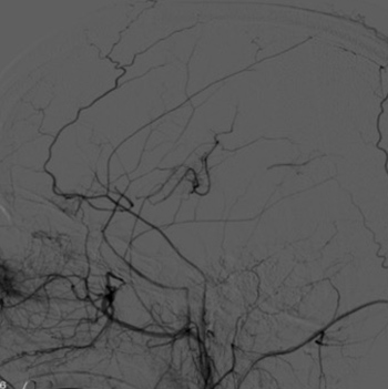
Moyamoya syndrome (MMS) is a rare, chronic and progressive vasculopathy with a characteristic angiographic pattern and well-recognized predisposing conditions, such as cranial therapeutic radiation. We report the case of a 36-year-old Caucasian female with a history of craniopharyngioma treated with whole-brain radiotherapy 20 years previously. She was admitted to the emergency department with disorientation and imperceptible speech lasting for 1 hour. Upon examination, she had slight motor aphasia, without sensory or motor deficits. However, the neurological deficits worsened on standing position. The computed tomography (CT) angiogram and transcranial Doppler ultrasonography revealed occlusion of the distal portion of the left internal carotid artery (ICA). Mechanical thrombectomy was attempted without success. Head CT was repeated, revealing left periventricular acute ischaemic stroke. The cerebral angiography showed total occlusion of the left ICA with an exuberant network of transdural collateral vessels, confirming MMS. The patient completed a functional rehabilitation program with progressive improvement of deficits and maintained a multidisciplinary follow-up. MMS is a serious late complication from cranial radiation therapy and a well-described risk factor for ischaemic stroke in younger patients. Therefore, early detection and prompt treatment are mandatory, as the consequences can be disastrous, such as cognitive and neurologic decline due to repeated strokes.
|
Views: 879
HTML: 94
PDF: 361
|
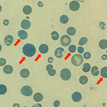
Introduction: Haemoglobin A1C (A1C), as a parameter of long-term glycaemic control, has been adopted to guide diabetic therapy all over the world. However, falsely high or low A1C could be troublesome in daily practice.
Case description: A 75-year-old male diabetic patient affected by a reasonably inferred life-long history of microcytic anaemia was found to have abnormally low A1C values in the previous 5 months. Subsequent laboratory assessment with brilliant cresyl blue staining and haemoglobin electrophoresis detected haemoglobin H disease as the underlying cause of both the microcytic anaemia and the disturbed A1C measurement.
Discussion: Enhanced erythrocyte destruction such as in haemoglobin H disease could explain a falsely decreased A1C level very well. Upon facing a questionable A1C value, physicians dealing with diabetes should consider the possibility of undiscovered underlying causes rather than too tightly glycaemic control.
|
Views: 960
HTML: 534
PDF: 319
|
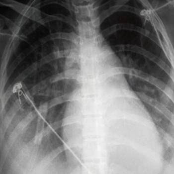
Introduction: Carbon monoxide (CO) poisoning may cause severe cellular hypoxia.
Materials and methods: A 28-year-old male presented reduced levels of consciousness and dyspnoea after CO exposure. Clinical examination revealed tachypnoea, bilateral rales, dilated jugular veins and confusion. Troponin I, lactate and carboxyhaemoglobin levels were increased. Thoracic X-ray depicted pulmonary oedema and an echocardiogram, severe heart failure (HF; EF<25%). He was intubated due to clinical deterioration.
Results: He remained intubated for 5 days with excellent improvement of left ventricular function (EF>55%). He was discharged 1 week later with full recovery.
Discussion: Acute HF is a rare serious complication of CO poisoning, even in healthy young individuals.
|
Views: 765
HTML: 77
PDF: 301
|
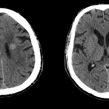
Aim: To describe an unusual presentation of a primary lymphoma of the central nervous system in a patient who, four months prior to admission, was diagnosed with herpes zoster ophthalmicus (HZO).
Case description: A 68-year-old man, with a history of HZO, was admitted to the emergency department with nausea and vomiting that had persisted over the previous two weeks. Neurological evaluation showed right ptosis, divergent strabismus and anisocoria. Blood tests showed high c-reactive protein, while serology was negative for human immunodeficiency virus. A brain CT scan revealed three round lesions, slightly hyperdense, periventricular in the occipital and frontal regions, which biopsy revealed to be a diffuse large B-cell lymphoma. The patient started chemotherapy but progression to death was inevitable.
Conclusion: The authors describe an unusual presentation of primary lymphoma of the central nervous system and urge physicians to be aware of this presentation in order to avoid misdiagnosis.
|
Views: 829
HTML: 212
PDF: 315
|
A woman in her early 40s, with a history of excessive alcohol intake, presented with purpuric, ulcerative lesions on the lower limbs. On examination, hirsutism and generalized stiffening and thickening of the skin were noted. Laboratory investigations revealed hyperbilirubinemia, hypergammaglobulinemia and positive anti-smooth muscle antibodies. Histologic examination of the skin was compatible with scleroderma. Histologic examination of the liver was suggestive of autoimmune and alcoholic hepatitis.
|
Views: 1086
HTML: 343
PDF: 574
|
Inclusion body myositis (IBM) is a chronic inflammatory myopathy with a progressive course. It is more common in the later years of life and usually presents with limb weakness. We present the case of a patient who developed proximal weakness in the lower limbs and, four years later, facial asymmetry. Blood analysis revealed high lactate dehydrogenase and creatinine kinase values. The diagnosis was obtained through muscle biopsy which met the histological criteria for IBM. The patient started treatment with alemtuzumab, leading to stabilisation of the symptoms in two years.

|
Views: 1349
HTML: 144
PDF: 360
|
Introduction:“Esophageal spasm” is a generic term widely used to attribute unexplained non-cardiac chest pain and/or dysphagia to an esophageal motility disorder.
Patient and methods: The authors present the case of an 86-year-old male patient with complete dysphagia after an elective electrical cardioversion for atrial fibrillation. An upper endoscopy performed shortly after the onset of the clinical picture documented disordered esophageal contractions. The patient became asymptomatic within 12 hours of the administration of a spasmolytic therapy.
Results: To the best of our knowledge, this is the first report of esophageal spasm after an electrical cardioversion.
Discussion: The temporal correlation supports the explanation of a cause-effect relationship between the clinical presentation and the preceding procedure, thus providing elements for clinicians to recognize and treat this particular condition.
| 2.1 = | 1.730 Cit. to date |
| 842 Docs. to date |


Publisher
Official Journal of the
European Federation of Internal Medicine
www.efim.org
Publisher: SMC media Srl
Via Giovenale, 7 - 20136 Milan - Italy
P.IVA 07626490960
info@ejcrim.com
www.ejcrim.com - ISSN: 2284-2594 - © EFIM 2014-2023, Published by SMC Media srl, Italy - Privacy policy

