EJCRIM 2023 CiteScore
| 2.1 = | 1.762 Cit. to date |
| 842 Docs. to date |
Last updated on 05 May, 2024
Updated monthly
Updated monthly
Powered by

|
Views: 1801
HTML: 103
PDF: 813
|
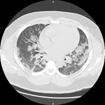
Background: Coronavirus disease 2019 (COVID-19) presents with a wide range of illness severity, from asymptomatic disease to severe acute respiratory distress syndrome (ARDS). Immunosuppression is considered a risk factor for severe COVID-19, but there are only few reports on disease progression in immunocompromised patients.
Case Summary: We report the case of a 50-year-old patient with acute COVID-19 pneumonia, who had iatrogenic, clinically relevant bone marrow suppression due to accidental overdose with hydroxyurea, and decreased lung capacity due to a left-sided pneumonectomy 6 months earlier. Symptomatic treatment with oxygen supplementation and pulmonary physical therapy was initiated, and hydroxyurea was discontinued. Over 14 days, the patient’s blood counts slowly recovered, and his clinical condition gradually improved, such that supplemental oxygen was no longer necessary and he could be discharged.
Discussion: A gradual increase in neutrophil and lymphocyte counts may be preferable to dampen a potentially detrimental immunological response triggered by severe acute respiratory syndrome coronavirus 2 (SARS-CoV-2). Whether patients with severe COVID-19 benefit from immunosuppressive therapy should be further evaluated.
|
Views: 1269
HTML: 87
PDF: 797
|
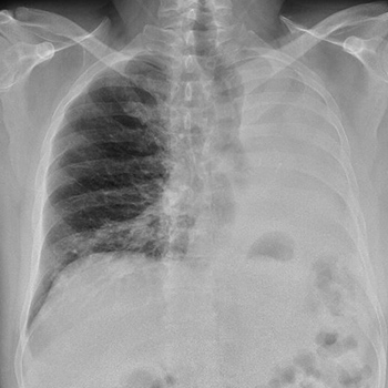
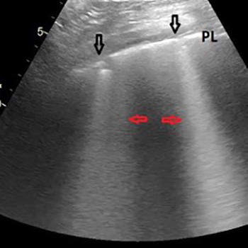
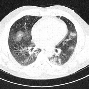
Coronavirus disease 2019 (COVID-19) is caused by severe acute respiratory syndrome coronavirus 2 (SARS-CoV-2) and the World Health Organization (WHO) declared it a pandemic on 11 March 2020. Point-of-care ultrasound (POCUS) is a real-time bedside tool used by physicians to guide rapid, focused and accurate evaluation in order to identify or rule out various pathologies. We describe the case of an elderly man who had fallen at home 3 days previously and was hypoxic at presentation to the emergency department (ED). POCUS in the ED helped to identify a combination of lung and vascular involvement that indicated COVID-19 infection, which was confirmed by a laboratory test.
|
Views: 1315
HTML: 197
PDF: 755
|
We describe a patient with SARS-CoV-2 and severe pneumonia who required mechanical ventilation and developed associated rhabdomyolysis with probable myocardial involvement as evidenced by cardiac enzyme abnormalities and echocardiographic findings. Repeat testing should be done in cases highly suspicious for SARS-CoV-2 as initial molecular tests may be negative, as in our case.
|
Views: 1827
HTML: 221
PDF: 892
|
Hydroxychloroquine has been used worldwide as a first-line treatment for patients hospitalized with COVID-19. Little is known about COVID-19 and its effects on patients with congenital red blood cell disorders. We report a case of haemolytic anaemia in a 32-year-old patient and a fortuitous highlighting of G6PD deficiency. We reviewed the literature to assess the risk of hydroxychloroquine use in this context.

|
Views: 995
HTML: 106
PDF: 576
|
Bacille Calmette–Guérin (BCG) administration for superficial bladder cancer is a well-tolerated and very effective therapy. However, unpredictable systemic complications may occur on rare occasions. We present the case of a patient who attended for consultation because of fever, asthenia and weight loss following BCG immunotherapy. The clinical response to treatment and computed tomography scanning were key to diagnosis.
|
Views: 1322
HTML: 79
PDF: 678
|
Acquired haemophilia A (AHA) is a rare autoimmune disorder caused by an autoantibody against any circulating coagulation factor, especially factor VIII (FVIII). The lack of awareness of this condition suggests that diagnosis is a challenge and usually delayed, which leads to suboptimal treatment. Consequently, early diagnosis is mandatory to prevent potentially life-threatening bleeding complications. We present the case of an 85-year-old woman admitted to hospital with symptoms of respiratory infection who 12 hours later developed haematuria which required transfusion. Laboratory assays showed an isolated prolonged aPTT, a moderately reduced FVIII and a high inhibitor titre. Influenza A and Escherichia coli were also identified. Antivirals, antibiotics, immunosuppressive drugs and haemostatic agents were started. Two weeks later, the inhibitor was not detected, and bleeding and symptoms of infection had resolved. Immunosuppressive drugs were stopped on day 45 and there has been no recurrence since then. To date, no FVIII inhibitors have been reported in concomitant infection with influenza A and urinary E. coli. The identification of conditions potentially associated with AHA is essential to achieve complete remission.
|
Views: 1543
HTML: 174
PDF: 862
|
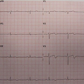
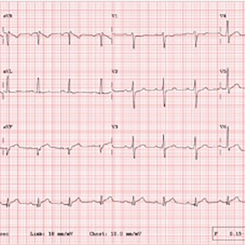
Wellens syndrome (WS) is identified by ECG changes in the precordial leads after resolution of angina chest pain. WS indicates critical stenosis of the proximal left anterior descending (LAD) artery. On the other hand, Kounis syndrome (KS) is an allergic reaction to various substances resulting in acute coronary syndrome. Contrast media can trigger the allergic reaction associated with KS. We describe a patient with WS who developed an allergic reaction to contrast media after percutaneous coronary intervention and experienced recurren myocardial infarction on re-exposure.
|
Views: 1894
HTML: 3486
PDF: 792
|
Non-alcoholic fatty liver disease (NAFLD) is the most common chronic liver disease and has emerged as a serious public health challenge. About 20% of NAFLD patients may have low titres (<1:320) of antinuclear antibodies (ANA). However, we describe a patient with NAFLD whose ANA titre was high (>1:320) on presentation. After 3 months of diet, exercise and vitamin E supplementation,the patient was symptomatically better but her ANA titre had increased (>1:640). Her liver biopsy showed features of NAFLD with minimal fibrosis. High-titre ANA (>1:320) positivity is rare. Our patient showed a progressive rise in ANA titre from >1:320 to >1:640 within 3 months even though she was improving and histology showed minimal fibrosis.
|
Views: 1073
HTML: 141
PDF: 687
|
Eribulin is an antineoplastic agent used in advanced breast cancer refractory to anthracycline and taxane treatment regimens. A wide variety of side effects with unclear mechanisms have been noted, but encephalopathy has not been widely reported. Here we describe the case of a middle-aged woman treated with eribulin for advanced breast cancer who subsequently developed central nervous system drug-induced toxicity but improved promptly with steroid administration.
|
Views: 970
HTML: 249
PDF: 643
|
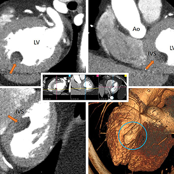
Cardiac lipomas are rare benign primary neoplasms of the heart, usually found incidentally, that can become symptomatic depending on their size and location. We report the case of a 61-year-old man presenting with chest pain and elevated troponin and a normal EKG and D-dimers. A transthoracic echocardiogram revealed an intracardiac mass attached to the interventricular septum protruding to the left ventricle, later confirmed to be a lipomatous mass consistent with a cardiac lipoma on cardiac magnetic resonance imaging. Due to the mass characteristics and favourable evolution, it was decided not to excise the tumour, and the patient remains asymptomatic after a 4-year follow-up period.
|
Views: 1370
HTML: 331
PDF: 549
|
Heparin is commonly used in clinical practice for the prevention and treatment of various thrombotic conditions. Its use can be associated with bleeding which can range from minor to life threatening. Non-traumatic causes of breast haematoma are very rare. We report a case of spontaneous bleeding into the breast in a female patient who was anticoagulated with heparin.
|
Views: 1193
HTML: 724
PDF: 706
|
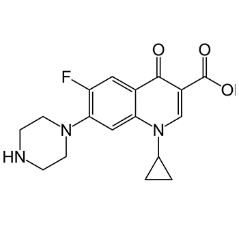
Fatal hepatotoxicity associated with ciprofloxacin is extremely rare. This is the second fully investigated case of fulminant hepatotoxicity due to ciprofloxacin in a male patient previously ciprofloxacin tolerant. The patient’s medical history included stable Waldenstrom’s macroglobulinaemia, inguinal hernia repair, prostate cancer (radiotherapy in 2006) and idiopathic Parkinson’s disease. Extensive investigation for progressive liver failure confirmed drug-induced liver injury.
|
Views: 902
HTML: 185
PDF: 566
|
Acquired haemophilia is a bleeding disorder caused by antibodies against coagulation factors. Some cases are associated with autoimmune diseases. However, no cases of acquired haemophilia with eosinophilic fasciitis have been previously reported. Herein we describe the case of a patient with eosinophilic fasciitis associated with acquired haemophilia.
|
Views: 1310
HTML: 127
PDF: 584
|
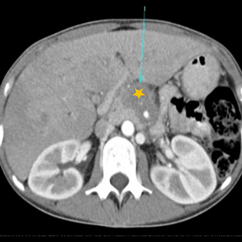
Inflammatory myofibroblastic tumour (IMT) is a rare mesenchymal tumour. It is usually benign but may behave as a malignant tumour with multiple recurrences and metastases. We present the case of a young woman with weight loss associated with diffuse abdominal pain, who was shown to have a large pancreatic mass. Investigation revealed fusocellular mesenchymal neoplasia, compatible with the diagnosis of IMT. As the mass was unresectable, glucocorticoid therapy was initiated with an excellent response and regression of the tumour.
|
Views: 998
HTML: 68
PDF: 507
|
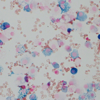
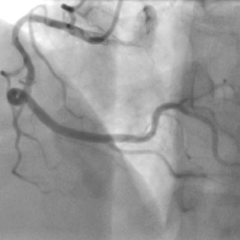
An 81-year-old man complaining of exertional dyspnoea underwent coronary angiography using an iodinated contrast medium. After angiography, the patient required systemic corticosteroid therapy because of respiratory failure due to alveolar haemorrhage. Percutaneous coronary intervention was performed 29 days after angiography using the same contrast medium. After the intervention, the patient required intubated mechanical ventilation and renal replacement therapy. Bronchoalveolar lavage was bloody with many haemosiderin-filled macrophages. Systemic corticosteroid therapy again improved his clinical condition. Iodinated contrast media may cause alveolar haemorrhage and re-exposure to contrast media may induce a more severe adverse reaction.
|
Views: 1160
HTML: 128
PDF: 598
|
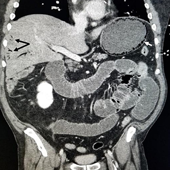
Body packing was first described in 1973 and refers to the intracorporeal concealment of illegal drugs, which are swallowed or placed in anatomical cavities and/or body orifices. The body packer can be asymptomatic or can have signs of systemic drug toxicity (neurological, cardiac, abdominal, renal and cutaneous) due to rupture of the packet(s) or symptoms of gastrointestinal obstruction or perforation. The diagnosis is established based on a suggestive history, findings on physical examination and laboratory findings and/or imaging. The vast majority of patients are asymptomatic and are treated conservatively. However, complex situations may require surgical intervention. We present a case of a 50-year-old man who was admitted in the emergency department with a generalized tonic-clonic seizure and vomiting with plastic film, which raised the suspicion of foreign body ingestion, confirmed by imaging and laboratory tests. He underwent exploratory laparotomy to remove the packages.
|
Views: 1308
HTML: 189
PDF: 623
|
Emphysematous gastritis is a rare but fatal variant of gastritis. It is caused by gastric wall invasion by gas-forming organisms. It follows disruption of gastric mucosal integrity by a variety of factors, most commonly caustic ingestion and alcohol abuse. Patients typically present with abdominal symptoms with features of septic shock. Emphysematous gastritis carries a high mortality rate warranting early intervention with supportive measures and broad-spectrum antibiotics. It is essential to consider this rare entity in the differential diagnosis of a patient presenting with abdominal pain as timely intervention is crucial for survival.
|
Views: 1211
HTML: 72
PDF: 656
|
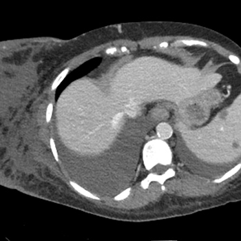
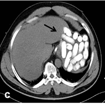
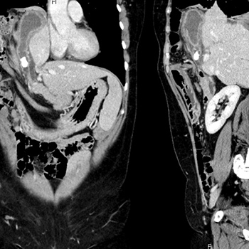
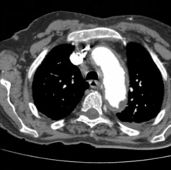
The authors present the case of a 51-year-old woman with no history of surgical or traumatic injury or accident, who presented with right hypochondrium and epigastric discomfort, malaise, nausea, loss of appetite and episodes of dark urine and greenish stools. Initial laboratory work-up revealed elevated inflammatory markers including leucocytosis with left shift and C-reactive protein, and a slight elevation of gamma-glutamyltransferase and alkaline phosphatase, with no other significant alterations. Computed tomography (CT) showed intrathoracic acute cholecystitis with a large diaphragmatic hernia.
A literature search revealed only one other case of acute cholecystitis complicated by intrathoracic gallbladder due to a non-traumatic diaphragmatic hernia. Symptoms are uncharacteristic and the absence of pain or fever, explained by the altered location of the gallbladder, makes the diagnosis a challenge.
|
Views: 998
HTML: 149
PDF: 903
|
Uterine leiomyomas are very common gynaecological benign tumours. Spontaneous torsion of a uterine subserosal leiomyoma is a rare cause of acute lower abdominal pain and should be treated immediately with surgery. We report a case of an enlarged subserosal leiomyoma that was first detected by computed tomography (CT) and further confirmed by laparoscopic surgery to be a subserosal leiomyoma with torsion.
|
Views: 1320
HTML: 521
PDF: 773
|
Objective: We present a case of a 22-year-old bodybuilder diagnosed with myocarditis secondary to clenbuterol use.
Results: The patient was primarily managed conservatively by the discontinuation of clenbuterol and the temporary use of dual anti-platelets, beta-blockers and nitrates.
Conclusion: Clenbuterol is a long-acting beta-2 agonist primarily used in veterinary medicine. In recent years, it has been illegally marketed as a weight loss supplement because of its anabolic properties and is popular among fitness enthusiasts. It is our aim to use this case to underscore the adverse effects of this drug with hopes that tighter regulations will be instituted to stem its illegal distribution.
|
Views: 1561
HTML: 148
PDF: 646
|
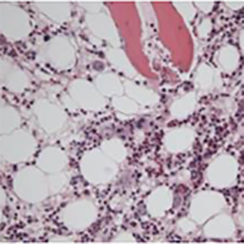
Reactivation of human parvovirus B19 is exceptional and characteristic of immunosuppression, with anaemia being the predominant manifestation although pancytopenia and thrombotic microangiopathy may also occur. We describe a patient with a history of diffuse large B-cell lymphoma with pure erythrocyte aplasia due to reactivation of parvovirus B19, who was treated with corticosteroids and immunoglobulins.
|
Views: 915
HTML: 248
PDF: 533
|
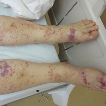
Diffuse dermal angiomatosis is a benign vascular disorder suspected in patients with cardiovascular risk factors. We report the case of a 62-year-old woman with a non-healing hip wound but no significant cardiovascular risk factors, who was found to have diffuse dermal angiomatosis on biopsy leading to the diagnosis of severe peripheral vascular disease. Her wound healed after revascularization.
|
Views: 844
HTML: 60
PDF: 598
|
We describe a case of coronary artery embolism leading to an out-of-hospital cardiac arrest (OHCA) in which the diagnosis was achieved with utilisation of cardiac magnetic resonance imaging. The patient was otherwise well prior to this episode. Emergency diagnostic coronary angiography revealed patent arteries with TIMI 3 flow. Subsequent cardiac magnetic resonance imaging demonstrated myocardial infarction and focal microvascular obstruction in the infarcted territory. This report describes an uncommon case presentation, highlights areas for improvement in diagnostic criteria, and briefly discusses the currently available data regarding coronary artery embolism.
|
Views: 1141
HTML: 221
PDF: 639
|
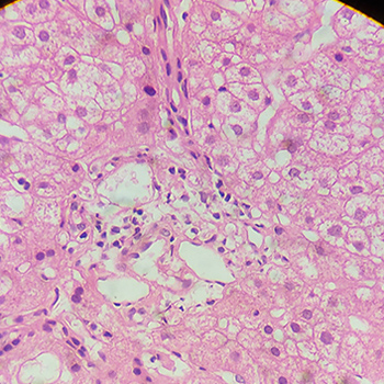
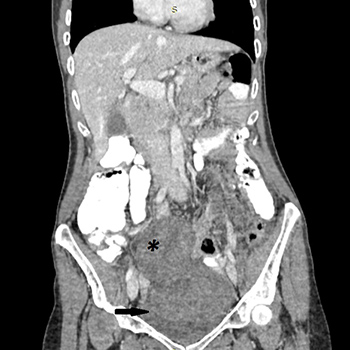
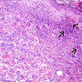
Pericardial effusion represents a diagnostic challenge. Erdheim-Chester disease (ECD), though a rare cause, should be considered in the differential diagnosis. An 88-year-old woman was admitted to the hospital due to retrosternal pain, dyspnoea and constitutional symptoms. Hypoxaemic respiratory failure and increased inflammatory markers were documented. A chest x-ray revealed an increased cardiothoracic ratio. An echocardiogram showed a moderate-volume pericardial effusion, without signs of cardiac tamponade. A thoraco-abdomino-pelvic CT scan found a bilateral perirenal soft tissue halo. Perirenal mass biopsy showed diffuse infiltration by foamy histiocytes (CD68+), without IgG4, compatible with ECD. The correlation of anamnesis, radiology and histology is crucial for the diagnosis of ECD.
|
Views: 998
HTML: 211
PDF: 605
|
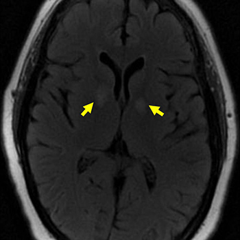
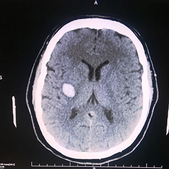
Spontaneous, non-traumatic intra-cerebral haemorrhage is the second most common type of stroke and is associated with significant morbidity and mortality. It is defined as the presence of blood within the cerebral parenchyma without prior injury or surgery. The purpose of this work is to describe an atypical presentation of spontaneous intra-cerebral haemorrhage in a healthy young adult. A literature review was also carried out.
| 2.1 = | 1.762 Cit. to date |
| 842 Docs. to date |


Publisher
Official Journal of the
European Federation of Internal Medicine
www.efim.org
Publisher: SMC media Srl
Via Giovenale, 7 - 20136 Milan - Italy
P.IVA 07626490960
info@ejcrim.com
www.ejcrim.com - ISSN: 2284-2594 - © EFIM 2014-2024, Published by SMC Media srl, Italy - Privacy policy

